Review
. 2008 Dec;4(6):815-24.
doi: 10.2217/14796694.4.6.815.

Fhit tumor suppressor: guardian of the preneoplastic genome

Affiliations
Review

Fhit tumor suppressor: guardian of the preneoplastic genome

Flavia Pichiorri et al. Future Oncol. 2008 Dec.

Abstract

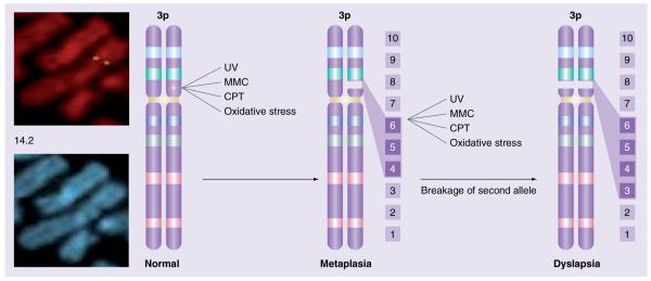
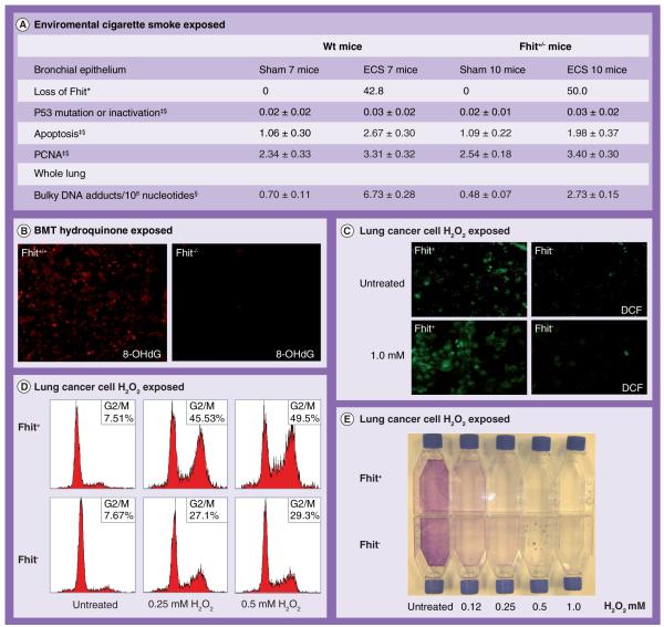
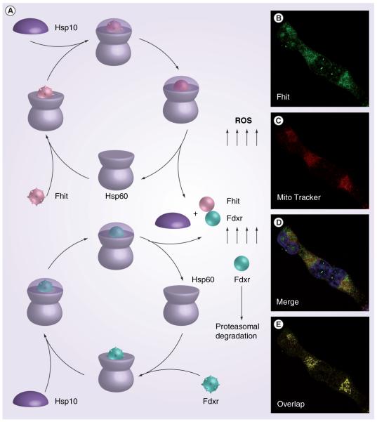
Environmental agents induce intragenic alterations in the FRA3B/FHIT chromosome fragile site, resulting in fragile FHIT allele loss early in cancer development. Fhit knockout mice are predisposed to tumor development and Fhit gene therapy reduces tumor burden. Repair-deficient cancers are likely to be Fhit-deficient and Fhit-deficient cells show enhanced resistance to ultraviolet C, mitomycin C, camptothecin and oxidative stress-induced cell killing. Loss of Fhit leads to alterations in the DNA damage response checkpoint and contributes to DNA instability. Hsp60/Hsp10 are Fhit interactors, suggesting a direct role for Fhit in stress responses. Fhit also interacts with and stabilizes ferrodoxin reductase (Fdxr), a mitochondrial flavoprotein that transfers electrons from NADPH to cytochrome P450, suggesting a role for Fhit in the modulation of reactive oxygen species production and of genomic damage.


Figures

Figure 1
Figure 1. Chromosome 3, showing the gap or break at 3p14 (right)
Hybridization of fluorescent genomic fragments of the 5′-end of the FHIT gene (green, left) shows the position of one end of FHIT flanking the fragile region. The carcinogens in cigarette smoke and other carcinogens also cause damage at FRA3B, leading to breakage of one FHIT allele with loss of exons 4–6, for example. Further carcinogen exposure can lead to damage at the second FHIT allele with loss of exons 3–5. Loss of one FHIT allele is frequently detected in the non-neoplastic epithelium of current and former smokers, and might lead to areas of metaplasia with reduced Fhit protein expression. Loss of the second FHIT allele can lead to total loss of Fhit protein expression (lower right), as observed in many dysplastic lesions. CPT: Cisplatin; MMC: Mitomycin C; UV: Ultraviolet.
Figure 2
Figure 2. Fhit in carcinogen-induced cancer and stress response
(A) Table summarizing alterations induced in B6–129(F1) mice, either wildtype or Fhit+/−, by whole-body exposure to ECS for 15 days [36]. *Percentage of mice showing extensive loss of Fhit within each experimental group; Percentage of mice with the reported alteration; §Means ± SE among all mice within each experimental group. (B) Detection of 8-OHdG representing the oxidative DNA damage in hydroquinone-exposed transplanted bone marrow cells; Fko and Wt bone marrow cells were exposed to hydroquinone and transplanted to recipient mice [38]. (C) Increased green fluorescent DCF signal in H1299 Fhit-expressing cells (D1) under stress condition [43]. (D) FACS ana lysis of D1 and E1 cell-cycle kinetics at 48 h after oxidative stress treatment; cells were treated with increasing concentrations of H2O2 (0.25, 0.5 mM) for 4 h. (E) Colony-formation assay of H1299/D1 (Fhit+) and H1299/E1 (Fhit) cells after 5 h treatment with H2O2 at indicated concentrations [43]. BMT: Bone marrow transplant; DCF: Dichlorofluorescein diacetate; ECS: Environmental cigarette smoke; PCNA: Proliferating cell nuclear antigen protein.
Figure 3
Figure 3. Model for the role of Fhit in protection of Fdxr from proteaosomal degradation
(A) Non-native Fhit (rough pink ball) and Fdxr (rough green ball) proteins bind to the trans ring of a Hsp60–Hsp10 complex. End-to-end exchange of Hsp10 results in the encapsulation of the protein substrate in the cis cavity. Release of Hsp10 and substrate proteins, Fhit and Fdxr, in the folded conformation (pink and green smooth balls, respectively) for mitochondrial addressing; we hypothesized that absence of Fhit leads to enhanced proteasomal degradation of Fdxr [43]. The immunofluorescence microscopy (B–E) was performed with antiFhit serum on H1299 Fhit-positive cells; Fhit staining was detected using fluorescein isothiocyanate (green) conjugated antirabbit immunoglobulin (IgG); MitoTracker Red staining, which identifies mitochondria, shows partial colocalization with Fhit. (E) The yellow color shows the colocalizations points.

References

    1. Croce CM. Role of chromosome translocations in human neoplasia. Cell. 1987;49(2):155–156. ▪▪ One of the most important reviews explaining the role of chromosome translocation and how they are related to cancer initiation.

    1. Soloman E, Borrow J, Goddart A. Chromosome aberrations and cancer. Science. 1991;254(5035):1153–1160. - PubMed
    1. Bartkova J, Horejsí Z, Koed K, et al. DNA damage response as a candidate anti-cancer barrier in early human tumorigenesis. Nature. 2005;434(7035):864–870. ▪▪ Reports an elegant study describing how cancer development is associated with DNA replication stress, which leads to DNA double-strand breaks, including breaks at FRA3B, checkpoint activation and selective pressure for p53 mutations.

    1. Gorgoulis VG, Vassiliou LV, Karakaidos P, et al. Activation of the DNA damage checkpoint and genomic instability in human precancerous lesions. Nature. 2005;434(7035):829–830. ▪▪ Reports an elegant study describing how cancer development is associated with DNA replication stress, which leads to DNA double-strand breaks, including breaks at FRA3B, checkpoint activation and selective pressure for p53 mutations.

    1. Naylor SL, Johnson BE, Minna JD, et al. Loss of heterozygosity of chromosome 3p markers in small cell lung cancer. Nature. 1987;329(6138):451–454. ▪ One of the first studies supporting the hypothesis that loss of alleles of chromosome 3p contributes to tumorigenesis in small-cell lung cancer.

Substances