. 2008 Dec 16:8:117.
doi: 10.1186/1471-213X-8-117.

Loss of Notch signalling induced by Dll4 causes arterial calibre reduction by increasing endothelial cell response to angiogenic stimuli

Affiliations

Loss of Notch signalling induced by Dll4 causes arterial calibre reduction by increasing endothelial cell response to angiogenic stimuli

Rui Benedito et al. BMC Dev Biol. .

Abstract

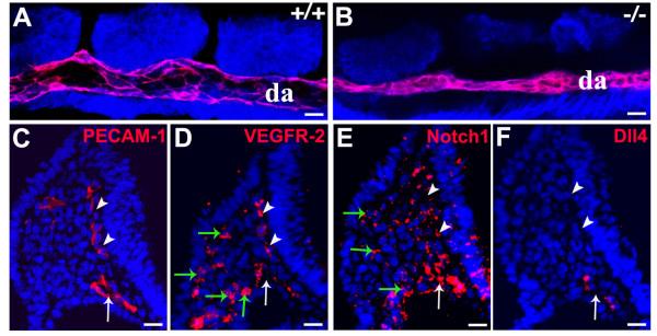
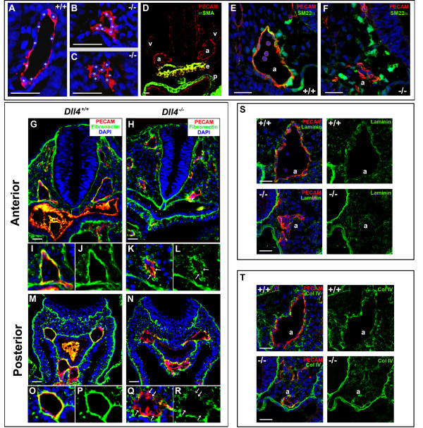
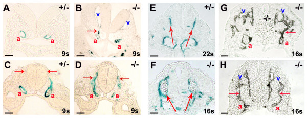
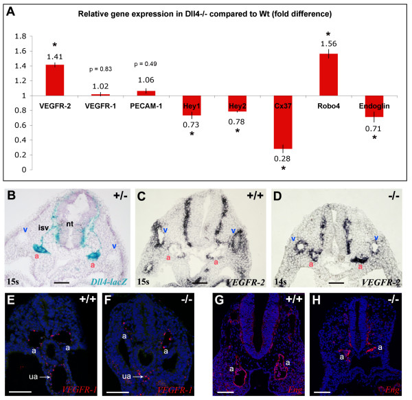
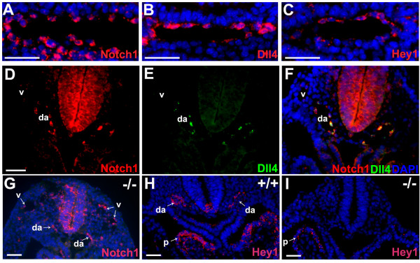
Background: In the vascular system, Notch receptors and ligands are expressed mainly on arteries, with Delta-like 4 (Dll4) being the only ligand known to be expressed early during the development of arterial endothelial cells and capillaries. Dll4 null embryos die very early in development with severely reduced arterial calibre and lumen and loss of arterial cell identity.

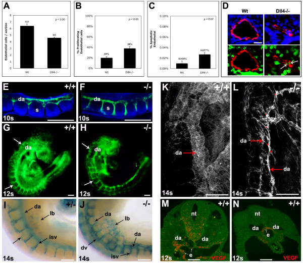
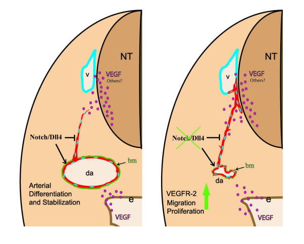
Results: The current detailed analysis of these mutants shows that the arterial defect precedes the initiation of blood flow and that the arterial Dll4-/- endothelial cells proliferate and migrate more actively. Dll4-/- mutants reveal a defective basement membrane around the forming aorta and increased endothelial cell migration from the dorsal aorta to peripheral regions, which constitute the main causes of arterial lumen reduction in these embryos. The increased proliferation and migration of Dll4-/- endothelial cells was found to coincide with increased expression of the receptors VEGFR-2 and Robo4 and with downregulation of the TGF-beta accessory receptor Endoglin.

Conclusion: Together, these results strongly suggest that Notch signalling can increase arterial stability and calibre by decreasing the response of arterial endothelial cells to local gradients of pro-angiogenic factors like VEGF.


Figures

Figure 1
Figure 1
The dorsal aortae calibre and lumen reduction in the Dll4-/- embryos is not caused by blood flow or angioblast defects. (A, B) Confocal imaging analysis of 8 ss Dll4+/+ and Dll4-/- embryos showing in red the dorsal aortae (da) endothelial cells (labelled with anti-PECAM-1) and in blue all the other cells from the adjacent tissues (labelled with anti-β-catenin). (C-F) Anti-PECAM-1 or fluorescent in situ hybridization on consecutive cryosections from a Dll4+/+ 8 ss embryo, showing that VEGFR-2 and Notch1 are expressed in the endothelial cells (PECAM-1+) and in the mesoderm cells from the lateral plate (green arrows), where angioblasts arise. The Dll4 mRNA is not detected on the angioblasts or on the primordial anterior cardinal vein (white arrowheads) but only on aortic endothelial cells (white arrows). (Scale Bars: 15 μm).
Figure 2
Figure 2
The Dll4-/- arterial endothelial cells have an abnormal morphology, become more aggregated and display a defective basement membrane. (A-C) Anti-PECAM-1 staining of 8 ss embryo cryosections showing the morphology of Dll4+/+ and Dll4-/- dorsal aortae endothelial cells. The white stars label the DAPI stained endothelial nuclei. Dll4-/- endothelial cells are more aggregated and fail to develop a stretched spindle-like form which leads to a reduced (B) or absent aortic lumen (C). (D) Anti-PECAM-1 (red) and anti-αSMA (green) staining of 16 ss cryosection of a Dll4+/+ embryo showing the absence of arterial (a) and venous (v) endothelium associated smooth muscle cells. Note the strong αSMA staining in the endoderm (e) and pericardium (p). (E, F) Both control and mutant dorsal aortae (a) are surrounded by neural crest derived smooth muscle cell progenitors with high amounts of Sm22α protein (in green). (G-R) Anti-PECAM-1 (red), anti-fibronectin (green) and DAPI (blue) staining of cryosections from the anterior (G-L) and posterior (M-R) region of 11 ss Dll4+/+ and Dll4-/- embryos. Figures I-L and O-R are close ups of the upper left dorsal aortae and show the defective basement membrane (green) in both the anterior and posterior regions of the Dll4-/- aortae. In the mutants many endothelial cells are not in direct contact with the underlying, fibronectin-rich, extracelular matrix (arrows). (S, T) At this stage of development the aortic (a) basement membrane is relatively poor in laminin (S, control) and collagenIV (T, control) when compared with fibronectin (J and P). The reduced Dll4-/- dorsal aortae have even less and more irregular laminin and collagenIV coverage. (Scale Bars: 30 μm).
Figure 3
Figure 3
Increased proliferation, sprouting and migration of Dll4-/- arterial endothelial cells. (A-C) Quantification of the endothelial cell number per aortic section (A), percentage of proliferating endothelial cells (B), and percentage of apoptotic endothelial cells (C) in the aortae of 8ss Wt and Dll4-/- embryos (n=2 for each genotype, 30 sections per embryo). (D) Representative images of the sections used for quantification. Endothelial cells are shown in red (anti-PECAM-1), nuclei in blue (DAPI) and proliferating cells in green (anti-BRDU). The proliferating endothelial cells are also indicated (arrows). (E-J) Whole-mount embryos labelled with anti-PECAM-1 (E-H, green) or X-gal (I, J) showing the increased migration of intersomitic vessel (isv) Dll4-/- endothelial cells. (E, F) Confocal imaging of the somitic region of 10ss embryos showing that in Dll4-/- embryos (F) the aortae endothelial cells invade the intersomitic space earlier and migrate extensively, causing the calibre reduction of the dorsal aortae (da). (G, H) At 12ss the endothelial cells start to accumulate in the dorsal domain (arrows) of the Dll4-/- embryos. (I, J) The X-gal staining of 14ss embryos shows the accumulation of endothelial cells (blue) in the dorsal region of the Dll4-/- first anterior-middle somites (J), forming a large dorsal vessel (dv). The Dll4-/- cells also migrate extensively to the primordial limb buds (lb). (K, L) Confocal imaging of 14ss wholemount embryos labelled with anti-PECAM-1. The Dll4-/- dorsal aortae (da) are thinner and show extensive sprouting (red dots). (M, N) In situ hybridization for VEGF mRNA on cryosections of a 12ss embryo shows that in the anterior region (M), VEGF is expressed by both the neural tube (nt) and endoderm (e) cells, but in the posterior (pre-somitic) region (N) is only expressed by the endoderm cells. Error bars indicate s.e.m. (Scale Bars: D, 25mm; E-J, 150mm; K-N, 100μm).
Figure 4
Figure 4
Dll4-/- endothelial cells migrate dorsally towards the neural tube, fusing with developing anterior cardinal veins. X-gal (A-F) and anti-PECAM-1 (G, H) staining of cryosections from the anterior (A, B) and middle (C-F) region of the Dll4+/- and Dll4-/- embryos with 8.5 (A-D) and 9.25 dpc (E-H). (A-D) Dll4-/- arterial endothelial cells migrate (arrows in B and D) towards the dorso-lateral region of the neural tube. In the anterior region of the Dll4 null embryos, the aortic (a, β-galactosidase+) cells migrate towards the developing anterior cardinal vein (v), before the onset of circulation (B). (E, F) At later stages, in the middle region of the embryo, this dorsal migration leads to the delocalization of the dorsal aortae. The main circulation in the mutant embryos occurs through two vessels that localize laterally (F) and not ventrally (E) to the neural tube. (G, H) In the anterior region of the later-stage Dll4-/- embryos the fusion (arrows) between the dorsal sprouting of the aortae (a) and the anterior cardinal veins (v) is evident. (Scale Bars: 50 μm).
Figure 5
Figure 5
Higher expression of VEGFR-2 and Robo-4 but lower expression of Endoglin in Dll4-/- embryos. (A) Relative quantitative gene expression in Dll4-/- embryos compared to Dll4+/+ (n = 4 per group) show increased expression of VEGFR-2 and Robo-4 and lower expression of Endoglin in the Dll4-/- mutants. Values were normalized to those of β-actin. (B-D) In situ hybridization on 9.0 dpc embryos cryosections show upregulation of VEGFR-2 specifically in Dll4-/- dorsal aortae (a). (B) In a thick section (50 μm) of a Dll4+/- embryo strong expression of Dll4 can be observed on the dorsal aortae and intersomitic vessels but no expression is detected on veins (v). (C) In a Dll4+/+ embryo VEGFR-2 is highly expressed in the veins and weakly expressed on the Dll4 expressing dorsal aortae. (D) In the absence of the arterial Dll4, VEGFR-2 is expressed at the same or at a higher level in the dorsal aortae compared to the anterior cardinal veins. (E, F) VEGFR1 (Flt1) in situ hybridization (in red) showing similar endothelial expression levels between control and mutant aortae (a) and umbilical arteries (ua). DAPI in blue. (G, H) Imunostaining for Endoglin (CD105) showing similar protein levels in endothelial cells of control and mutant embryos. Error bars represent st.dev. * indicates p < 0.05. (Scale Bars: 100 μm).
Figure 6
Figure 6
Notch1 and Dll4 are expressed at different levels in different cells but at high levels in the same cell of the developing arteries. (A-C) In situ hybridization on consecutive cryosections of 8.75 dpc (12 ss) Dll4+/+ embryos showing that Notch1, Dll4 and Hey1 are expressed (red) in almost all aortic endothelial cells. (D-F) Double in situ hybridization for Notch1 and Dll4 reveals that these genes are expressed at high levels in the same cells. Note also that Notch1 is weakly expressed on the anterior cardinal veins (v), where there is no expression of Dll4. (G-I) In the Dll4-/- embryos the Notch1 receptor is still expressed on dorsal aortae (da) but there is no detectable endothelial expression of the effector Hey1 (I), although its expression remains in the pericardium (p). (Scale Bars: 50 μm).
Figure 7
Figure 7
Regulation of arterial calibre and endothelial cell migration by Dll4. Model illustrating the difference in the behaviour of endothelial cells in the presence or absence of Notch signalling mediated by the Dll4 ligand in the early embryo arterial endothelium. The loss of Notch signalling leads to downregulation of arterial specific genes and upregulation of VEGFR-2 which may increase migration and proliferation of endothelial cells. The dorsal aortae (da) endothelial cells migrate towards the lateral region of the neural tube (nt) or pre-formed veins (v) with which they fuse. In the Dll4+/+ embryos Notch signalling induces arterial endothelial cell differentiation and stabilization, as well as promoting the formation of a consistent basement membrane (bm).

References

    1. Risau W. Mechanisms of angiogenesis. Nature. 1997;386:671–4. doi: 10.1038/386671a0. - DOI - PubMed
    1. Carmeliet P, Ferreira V, Breier G, Pollefeyt S, Kieckens L, Gertsenstein M, Fahrig M, Vandenhoeck A, Harpal K, Eberhardt C, et al. Abnormal blood vessel development and lethality in embryos lacking a single VEGF allele. Nature. 1996;380:435–9. doi: 10.1038/380435a0. - DOI - PubMed
    1. Shalaby F, Rossant J, Yamaguchi TP, Gertsenstein M, Wu XF, Breitman ML, Schuh AC. Failure of blood-island formation and vasculogenesis in Flk-1-deficient mice. Nature. 1995;376:62–6. doi: 10.1038/376062a0. - DOI - PubMed
    1. Fong GH, Rossant J, Gertsenstein M, Breitman ML. Role of the Flt-1 receptor tyrosine kinase in regulating the assembly of vascular endothelium. Nature. 1995;376:66–70. doi: 10.1038/376066a0. - DOI - PubMed
    1. Fong GH, Zhang L, Bryce DM, Peng J. Increased hemangioblast commitment, not vascular disorganization, is the primary defect in flt-1 knock-out mice. Development. 1999;126:3015–25. - PubMed

Publication types

MeSH terms

LinkOut - more resources