. 2008 Dec 15;14(24):8228-35.
doi: 10.1158/1078-0432.CCR-08-1329.

The incidence, correlation with tumor-infiltrating inflammation, and prognosis of phosphorylated STAT3 expression in human gliomas

Affiliations

The incidence, correlation with tumor-infiltrating inflammation, and prognosis of phosphorylated STAT3 expression in human gliomas

Mohamed Abou-Ghazal et al. Clin Cancer Res. .

Abstract

Purpose: The signal transducer and activator of transcription 3 (STAT3) is frequently overexpressed in most cancers, propagates tumorigenesis, and is a key regulator of immune suppression in cancer patients. We sought to determine the incidence of phosphorylated STAT3 (p-STAT3) expression in malignant gliomas of different pathologic types, whether p-STAT3 expression is a negative prognostic factor, and whether p-STAT3 expression influences the inflammatory response within gliomas.

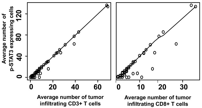
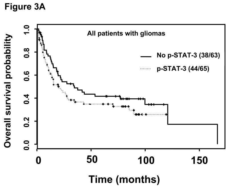
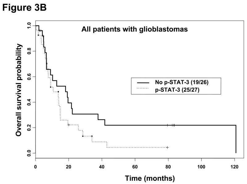
Methods: Using immunohistochemical analysis, we measured the incidence of p-STAT3 expression in 129 patients with gliomas of various pathologic types in a glioma tissue microarray. We categorized our results according to the total number of p-STAT3-expressing cells within the gliomas and correlated this number with the number of infiltrating T cells and T regulatory cells. We then evaluated the association between p-STAT3 expression and median survival time using univariate and multivariate analyses.

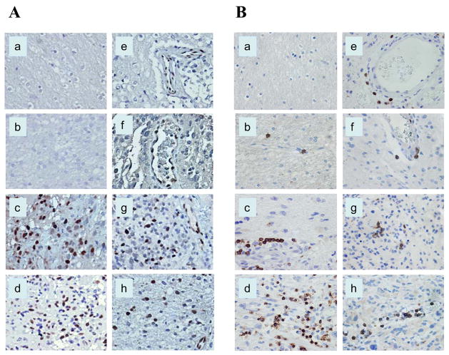
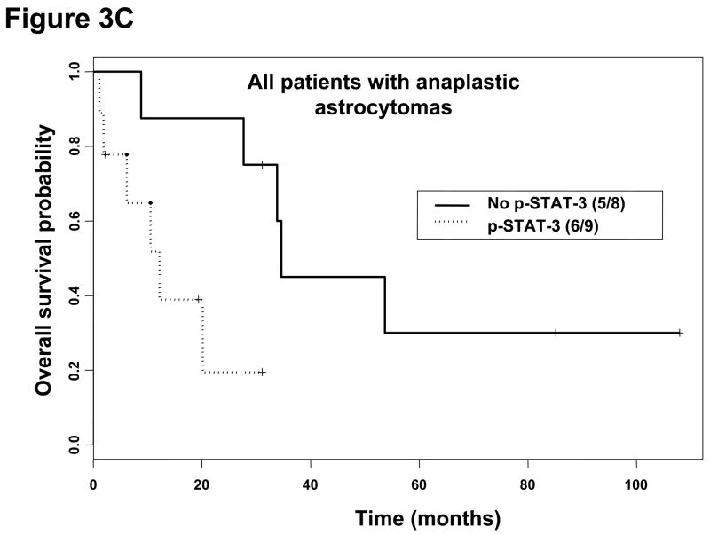
Results: We did not detect p-STAT3 expression in normal brain tissues or low-grade astrocytomas. We observed significant differences in the incidence of p-STAT3 expression between the different grades of astrocytomas and different pathologic glioma types. p-STAT3 expression was associated with the population of tumor-infiltrating immune cells but not with that of T regulatory cells. On univariate analysis, we found that p-STAT3 expression within anaplastic astrocytomas was a negative prognostic factor.

Conclusions: p-STAT3 expression is common within gliomas of both the astrocytic and oligodendroglial lineages and portends poor survival in patients with anaplastic astrocytomas. p-STAT3 expression differs significantly between gliomas of different pathologic types and grades and correlated with the degree of immune infiltration.


Figures

Figure 1
Figure 1
Immunohistochemical staining of human glioma tissue sections demonstrating p-STAT3 and CD3+ lymphoid cells. The p-STAT3 staining was confined to the nucleus, whereas CD3 staining was noted on the cell surface. A, The number of p-STAT3–expressing cells was more evident in astrocytic, higher-grade gliomas. B, CD3 staining demonstrated high numbers of infiltrative CD3+ T cells, also within higher-glioma grades. All images were taken at a magnification of x400. Normal brain (a), low-grade astrocytoma (b), anaplastic astrocytoma (c), glioblastoma multiforme (d), oligodendroglioma (e), mixed oligoastrocytoma (f), anaplastic oligodendroglioma (g), and anaplastic mixed oligoastrocytoma (h).
Figure 2
Figure 2
Pair-wise scatter plots between p-STAT3, CD3, and CD8 across all tumor pathologies and tumor grades with Loess smooth curves added. The almost straight trend of the Loess curves indicates that both CD3 and CD8 had a strong linear correlation with p-STAT3 expression. Specifically, the correlation of the number of p-STAT3 expressing cells with the number of tumor infiltrating CD3+ T cells was 0.99 (P < 0.001) and with the number of tumor infiltrating CD8+ T cells was 0.94 (P < 0.001).
Figure 3
Figure 3
Kaplan-Meier survival estimates stratified by the presence or absence of p-STAT3 immunohistochemical staining within all patients with gliomas, patients with glioblastoma multiforme, and patients with anaplastic astrocytoma. A, Among all patients with gliomas without p-STAT3 expression, the median survival time was 34.6 months (95% CI, 19.2, months-A) and 20.1 months (95% CI, 13.8–43.0 months) in patients with gliomas with p-STAT3 expression (P = 0.16). B, In patients with glioblastoma multiforme without p-STAT3 expression, the median survival time was 18.1 months (95% CI, 6.6–41.8 months) and 10.7 months (95% CI, 7.5–15.0 months) in patients with glioblastoma multiforme with p-STAT3 expression (P = 0.12). C, In patients with anaplastic astrocytoma patients without p-STAT3 expression, the median survival time was 34.6 months (95% CI, 33.9 months-NA) and 12.2 months (95% CI, 6.2 months-NA) in patients with anaplastic astrocytoma with p-STAT3 expression (P = 0.02).
Figure 3
Figure 3
Kaplan-Meier survival estimates stratified by the presence or absence of p-STAT3 immunohistochemical staining within all patients with gliomas, patients with glioblastoma multiforme, and patients with anaplastic astrocytoma. A, Among all patients with gliomas without p-STAT3 expression, the median survival time was 34.6 months (95% CI, 19.2, months-A) and 20.1 months (95% CI, 13.8–43.0 months) in patients with gliomas with p-STAT3 expression (P = 0.16). B, In patients with glioblastoma multiforme without p-STAT3 expression, the median survival time was 18.1 months (95% CI, 6.6–41.8 months) and 10.7 months (95% CI, 7.5–15.0 months) in patients with glioblastoma multiforme with p-STAT3 expression (P = 0.12). C, In patients with anaplastic astrocytoma patients without p-STAT3 expression, the median survival time was 34.6 months (95% CI, 33.9 months-NA) and 12.2 months (95% CI, 6.2 months-NA) in patients with anaplastic astrocytoma with p-STAT3 expression (P = 0.02).

References

    1. Masamune A, Satoh M, Kikuta K, Suzuki N, Shimosegawa T. Activation of JAK-STAT pathway is required for platelet-derived growth factor-induced proliferation of pancreatic stellate cells. World J Gastroenterol. 2005;11:3385–91. - PMC - PubMed
    1. Huang S. Regulation of metastases by signal transducer and activator of transcription 3 signaling pathway: clinical implications. Clin Cancer Res. 2007;13:1362–6. - PubMed
    1. Yu H, Jove R. The STATs of cancer--new molecular targets come of age. Nat Rev Cancer. 2004;4:97–105. - PubMed
    1. Li B, Chang CM, Yuan M, McKenna WG, Shu HK. Resistance to small molecule inhibitors of epidermal growth factor receptor in malignant gliomas. Cancer Res. 2003;63:7443–50. - PubMed
    1. Tarkowski E, Rosengren L, Blomstrand C, et al. Early intrathecal production of interleukin-6 predicts the size of brain lesion in stroke. Stroke. 1995;26:1393–8. - PubMed

Publication types

Substances