. 2009 Jan;136(2):275-83.
doi: 10.1242/dev.019042. Epub 2008 Dec 15.

The Bax/Bak ortholog in Drosophila, Debcl, exerts limited control over programmed cell death

Affiliations

The Bax/Bak ortholog in Drosophila, Debcl, exerts limited control over programmed cell death

Kathleen A Galindo et al. Development. 2009 Jan.

Abstract

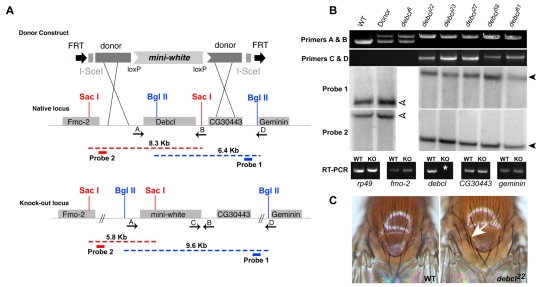
Bcl-2 family members are pivotal regulators of programmed cell death (PCD). In mammals, pro-apoptotic Bcl-2 family members initiate early apoptotic signals by causing the release of cytochrome c from the mitochondria, a step necessary for the initiation of the caspase cascade. Worms and flies do not show a requirement for cytochrome c during apoptosis, but both model systems express pro- and anti-apoptotic Bcl-2 family members. Drosophila encodes two Bcl-2 family members, Debcl (pro-apoptotic) and Buffy (anti-apoptotic). To understand the role of Debcl in Drosophila apoptosis, we produced authentic null alleles at this locus. Although gross development and lifespans were unaffected, we found that Debcl was required for pruning cells in the developing central nervous system. debcl genetically interacted with the ced-4/Apaf1 counterpart dark, but was not required for killing by RHG (Reaper, Hid, Grim) proteins. We found that debcl(KO) mutants were unaffected for mitochondrial density or volume but, surprisingly, in a model of caspase-independent cell death, heterologous killing by murine Bax required debcl to exert its pro-apoptotic activity. Therefore, although debcl functions as a limited effector of PCD during normal Drosophila development, it can be effectively recruited for killing by mammalian members of the Bcl-2 gene family.


Figures

Fig. 1.
Fig. 1.
Generation and verification of a targeted debcl mutation. (A) Targeting scheme for the debcl gene. The donor construct was generated by insertional cloning of a 4 kb upstream and a 2.7 kb downstream genomic sequence of the debcl gene into the targeting vector pW25. Upon chromosomal targeting, the debcl native gene is replaced with a white+ marker gene. (B) Verification of the debcl-targeting event by PCR, Southern blot and RT-PCR analysis. Primer pair A and B were used to amplify both the native and the knock-out locus to confirm the replacement of debcl (4.2 kb) with the white+ marker gene (∼5.2 kb). Primer pair C and D were used to verify the right arm of recombination during the screening process for potential recombinants. Primer pair E and F were used to confirm targeted recombination of the left arm of recombination (data not shown). WT refers to the yw parental strain, wild type at the debcl locus on the second chromosome; Donor refers to the debcl donor construct on the third chromosome, wild-type at the debcl native locus on the second chromosome; debcl6,22,23,27,59,61 represent different debcl knock-out candidates disrupted at the native locus. debcl6 was a potential debcl targeted deletion, but failed the PCR screen and was therefore eliminated from further characterization analysis. An additional debcl disruption allele, debcl47/48, was also confirmed (not shown). Southern blot analysis using genomic DNA from the indicated fly strains was used to confirm both the right (SBP1) and left (SBP2) arm of recombination. For SBP1 (BglII digest) and SBP2 (SacI digest), the black arrowheads indicate the expected 6.4 kb and 8.3 kb genomic fragments in the WT and donor strains for SBP1 and SBP2, respectively. The white arrowheads indicate aberrant genomic fragments of 9.6 kb and 5.8 kb, indicative of gene-targeted replacement, for SBP1 and SBP2, respectively. RNA from L3 larvae was used as a template to confirm abolishment of the debcl transcript in debclKO flies, and to confirm that transcript levels are unaffected in the neighboring genes fmo-2, CG30443 and geminin. rp49 was used as a control for RT-PCR. The white asterisk indicates the absence of the debcl transcript in mutant flies. (C) Photograph of extra scutellar bristles in debclKO flies (white arrow).
Fig. 2.
Fig. 2.
debcl regulates developmental cell death in the nervous system. (A,B) TUNEL staining is moderately reduced in debcl mutant embryos (B) compared with WT (yw) embryos (A; stage 11). (C-H) The presence of extra cells during embryonic development was examined by α-Kr Ab staining (C-G) and slit-lacZ staining (I,J slit-lacZ reporter; K,L, α-βGal Ab staining). Arrows in J-L indicate extra slit-lacZ+ cells. Dashed lines in E and H represent the WT cell count threshold for each segment surveyed. (E) Quantification of extra cells in the VNC by α-Kr Ab. Error bars show ±s.d. (n=6). (H) Quantification of cells in Bolwig's Organ. Error bars show ±s.d. (n=8). (M) Quantification of VNC cells by α-βGal Ab staining. Error bars show ±s.d. (n=8).
Fig. 3.
Fig. 3.
debcl and genotoxic stress. (A-N) After irradiation, debcl mutants show an elevated incidence of visible defects, but are unaffected for stress-induced apoptosis and cell-cycle arrest. (A,B) Basal levels of cell death are observed in the larval wing imaginal disc. (C,D) Four hours after exposure to 4000 Rads of ionizing radiation (IR), robust cell death is detected by Acridine Orange staining (AO). (M) Adult survival curve after treatment with IR. The corresponding LD50 is shown for each sample (dashed line). Effector caspase levels labeled by α-proCaspase3 Ab staining (G) drastically increased after IR challenge in WT and debcl22/27 tissue (J, WT not shown). Cell proliferation in the larval L3 wing imaginal disc was examined before (H,I) and after (K,L) treatment with 4000 Rads of IR, by α-phosphohistone H3 Ab staining. Wing from a WT (yw, E) and debcl22/27 (F) adult after IR challenge, showing a notched wing phenotype in the debcl mutant. (N) Histogram quantifying the percentage of notched wings in WT (yw) and transheterozygous debcl mutant alleles at 0, 1500 and 2500 Rads of IR. Dashed line represents WT percentage of notched wings.
Fig. 4.
Fig. 4.
debcl genetically interacts with dark. (A-C) Adult wings are normal in morphology and appearance at the time of eclosion (D0). (D-F) Wing blemishes are progressive in nature and more severe in debcl, darkCD4 double mutants (E,F) than in darkCD4 single mutants (D), seen here in 14-day-old animals. (G) Histogram quantifying the severity of wing blemishing. The incidence of wing blemishing is expressed as a percentage of the wing afflicted with melanized spots over the entire wing region. Error bars show ±s.d. (n=10).
Fig. 5.
Fig. 5.
Ectopic expression of pro-apoptotic genes in debcl mutant animals. (A,B) A morphologically normal eye in WT (A) and debcl59 (B). Killing by the RHG proteins is not dependent on debcl function. (C-H) pGMR-rpr (C,D), pGMR-grim4 (E,F) and pGMR-hid (G,H) in the eye showing no effect on the ablated eye phenotype. (I,K) Two different alleles of UAS-Bax1, GMR-Gal4 showing severe ablation of the eye in the WT background. (J,L) This severe eye phenotype is partially restored in a debcl heterozygous background (not shown) and is nearly completely reversed in homozygous debclKO; UAS-Bax1, GMR-Gal4.
Fig. 6.
Fig. 6.
debcl mutants display normal starvation-induced autophagy. (A) Histogram illustrating the survival curve of L2 larvae when placed under starved conditions (20% sucrose). (B) Fat bodies of young L3 larvae were quantified for autophagic bodies under fed (wet yeast) and starved (20% sucrose) conditions using lysotracker as a marker for autophagy. Error bars show ± s.d. (n=5). (C-F) Live fat bodies stained with Hoechst (blue) and lysotracker (green) in fed and starved larvae of wild type and mutant.
Fig. 7.
Fig. 7.
debcl and mitochondrial dynamics. (A-D) Images of WT (A,C) and debclKO (B,D) mitochondria (UAS-mito-GFP; Da-Gal4) in cells of L3 wing imaginal discs and salivary glands. Mitochondria from pupal salivary glands ∼13 hours after puparium formation (APF) are fragmented in response to PCD in WT and debclKO cells (not shown). (E-H) Mitochondrial density (E,F) and volume (G,H) in both tissues is comparable.

References

    1. Abdelwahid, E., Yokokura, T., Krieser, R. J., Balasundaram, S., Fowle, W. H. and White, K. (2007). Mitochondrial disruption in Drosophila apoptosis. Dev. Cell 12, 793-806. - PubMed
    1. Arama, E., Agapite, J. and Steller, H. (2003). Caspase activity and a specific cytochrome C are required for sperm differentiation in Drosophila. Dev. Cell 4, 687-697. - PubMed
    1. Arama, E., Bader, M., Srivastava, M., Bergmann, A. and Steller, H. (2005). The two Drosophila cytochrome C proteins can function in both respiration and caspase activation. EMBO J. 25, 232-243. - PMC - PubMed
    1. Brachmann, C. B., Jassim, O. W., Wachsmuth, B. D. and Cagan, R. L. (2000). The Drosophila Bcl-2 family member dBorg-1 functions in the apoptotic response to UV-irradiation. Curr. Biol. 10, 547-550. - PubMed
    1. Chen, P. and Abrams, J. M. (2000). Drosophila apoptosis and Bcl-2 genes: Outliers fly in. J. Cell Biol. 148, 625-627. - PMC - PubMed

Publication types

MeSH terms