. 2009 Mar 1;18(5):919-30.
doi: 10.1093/hmg/ddn430. Epub 2008 Dec 16.

Biallelic somatic and germline mutations in cerebral cavernous malformations (CCMs): evidence for a two-hit mechanism of CCM pathogenesis

Affiliations

Biallelic somatic and germline mutations in cerebral cavernous malformations (CCMs): evidence for a two-hit mechanism of CCM pathogenesis

Amy L Akers et al. Hum Mol Genet. .

Abstract

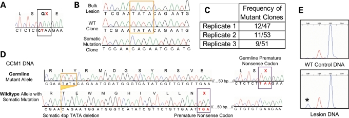
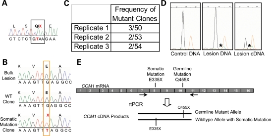
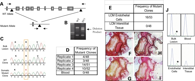
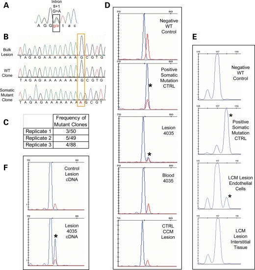
Cerebral cavernous malformations (CCMs) are vascular anomalies of the central nervous system, comprising dilated blood-filled capillaries lacking structural support. The lesions are prone to rupture, resulting in seizures or hemorrhagic stroke. CCM can occur sporadically, manifesting as solitary lesions, but also in families, where multiple lesions generally occur. Familial cases follow autosomal-dominant inheritance due to mutations in one of three genes, CCM1/KRIT1, CCM2/malcavernin or CCM3/PDCD10. The difference in lesion burden between familial and sporadic CCM, combined with limited molecular data, suggests that CCM pathogenesis may follow a two-hit molecular mechanism, similar to that seen for tumor suppressor genes. In this study, we investigate the two-hit hypothesis for CCM pathogenesis. Through repeated cycles of amplification, subcloning and sequencing of multiple clones per amplicon, we identify somatic mutations that are otherwise invisible by direct sequencing of the bulk amplicon. Biallelic germline and somatic mutations were identified in CCM lesions from all three forms of inherited CCMs. The somatic mutations are found only in a subset of the endothelial cells lining the cavernous vessels and not in interstitial lesion cells. These data suggest that CCM lesion genesis requires complete loss of function for one of the CCM genes. Although widely expressed in the different cell types of the brain, these data also suggest a unique role for the CCM proteins in endothelial cell biology.


Figures

Figure 1.
Figure 1.
A biallelic somatic mutation identified in CCM1 sample 2049. (A) Direct sequence analysis of bulk lesion tissue identified the germline mutation as the common Hispanic mutation, c.1363C>T, Q455X in exon 10 of CCM1. (B) Clonal analysis of CCM1 exon 10 reveals wild-type clones, germline mutant clones and clones with the 4 bp deletion somatic mutation, c.1270_1273DelTATA. The deleted bases are boxed in orange in the wild-type sequences, and the deletion site is marked with an orange dotted line in the somatic mutant sequence (bottom tracing). The somatic mutation is barely visible from direct sequence of bulk lesion. (C) The somatic mutation was identified in the lesion tissue multiple times in each of three rounds of PCR, cloning and sequencing. Replicates 1 and 2 used HiFi Platinum Taq Polymerase (Invitrogen) and replicate 3 used HotStar Polymerase (Qiagen). (D) The somatic and germline mutations are biallelic. Sequencing analysis of individual colonies containing both mutation loci shows three classes of clones, germline mutant alleles, wild-type alleles, and alleles carrying the somatic mutation that are wild-type for the germline mutation. (E) Fragment analysis validates the somatic mutation in the lesion. Normal control DNA shows all amplicons of predicted 252 bp length (blue peak). The red size marker is at 250 bp, and the lesion-derived DNA shows products of WT length and those from the somatic mutant allele at 248 bp. The mutant allele represents 11% of the total amplicons.
Figure 2.
Figure 2.
A biallelic somatic mutation identified in CCM1 sample 2009. (A) Direct sequence analysis of bulk lesion-derived DNA identified the germline mutation as the common Hispanic mutation, CCM1 exon 10 c.1363C>T, Q455X. (B) Somatic mutation analysis identified a somatic mutation in CCM1 exon 8, c1003G>T, E335X. Direct sequence of lesion DNA shows only wild-type sequence. Sequence of individual clones reveals sequence for both wild-type clones and clones with the somatic mutation. (C) The somatic mutation was identified multiple times in each of the three rounds of PCR. Replicates 1 and 2 used HiFi Platinum Taq Polymerase (Invitrogen), and replicate 3 used HotStar Polymerase (Qiagen). (D) The somatic mutation is validated using the SNapShot Assay. The wild-type allele in blue (arrow) is present in all samples, whereas the somatic mutation in green (asterisk) is only present in the lesion-derived DNA or cDNA products and orange peaks are the size standard. Somatic mutant alleles represent ∼6% of the DNA-derived fragments and 24% of the cDNA-derived fragments analyzed. (E) The somatic and germline mutations are biallelic. RT–PCR products containing both somatic and germline mutant loci were amplified, cloned and sequenced. Sequencing analysis of individual colonies shows three classes of clones: germline mutant alleles, wild-type alleles and alleles carrying the somatic mutation that are wild-type for the germline mutation.
Figure 3.
Figure 3.
A biallelic somatic mutation is identified in CCM2 sample 3911. (A) The germline mutation is identified as a large deletion of the CCM2 gene encompassing exons 2–10, which is readily screened for using primers spanning the deleted region (arrows). (B) Sample 3911 is heterozygous for the CCM2 2-10 deletion. The intermediate size band results from a PCR, which spans the 77 kb deletion (32), and is not present in wild-type controls. The upper and lower PCR products are control reactions, each requiring one of the primers used to generate the deletion product band (32). (C) Somatic mutation analysis reveals a somatic mutation, c.55C>T, R19X in exon 2 of the CCM2 gene. The somatic mutation is not readily apparent on a sequence tracing from bulk lesion. Sequence from multiple clones containing exon 2 inserts reveal both wild-type and somatic mutant clones. (D) The somatic mutation is present in multiple clones in each of three PCR replicates from bulk lesion tissue. Using template DNA derived from histology slides, the third replicate also had multiple clones with the somatic mutation. A different polymerase, Hotstar polymerase (Qiagen), was used for the final replicate. The somatic mutation is not present in clones generated from leukocyte-derived template DNA. (E) Laser capture microscopy was preformed to isolate endothelial cells surrounding the caverns as well as interstitial (non-endothelial) lesion tissue. The somatic mutation was identified using endothelial cell-derived DNA as template. The interstitial tissue-derived DNA clones were entirely wild-type. (F–H) Laser capture microscopy to isolate inter-cavernous control cells [(F) before LCM with targeted tissue outlined in black and (G) after isolation] and endothelial cells surrounding blood-filled caverns [(H) before LCM with targeted EC's outlined in black and (I) after LCM]. (J) The somatic mutation is validated using the SNapShot assay. The somatic mutation is present in bulk lesion and laser-captured endothelial cell-derived DNA (green peak with asterisks). The wild-type allele (blue) is present in all samples.
Figure 4.
Figure 4.
A CCM3 somatic mutation identified in sample 283-4035. (A) The germline mutation identified by sequence analysis of leukocyte-derived DNA is a splicing mutation in CCM3 exon 8, c.474+1 G>A. (B) Somatic mutation identified as an insertion of A in exon 6 of CCM3, c.205-211insA. The somatic mutation is not readily identifiable from the direct sequence of bulk lesion-derived DNA. Clonal analysis reveals both wild-type and somatic mutant clones. (C) The somatic mutation is identified multiple times in each of three replicates. The final replicate is of Phusion-derived PCR products. (D) The somatic mutation is validated by fragment analysis. Size standard marker at 75 bp is in red. Phusion-derived PCR fragments appear in blue. The wild-type fragment product is 74 bp (Negative Ctrl), and the somatic mutation product is 75 bp long (Positive Ctrl). The lesion-derived sample yields two products, both wild-type and mutant (asterisk) with the mutant allele representing 10% of the total amplicons. The larger fragment of the somatic mutation is not present in either matched blood for lesion sample 4035 or another unrelated CCM lesion sample that is WT at the site of the somatic mutation. (E) In a different fragment analysis assay, normal WT length fragments are 117 bp, and those with the somatic mutation (asterisk) are 118 bp long. From the laser-captured tissue, only the endothelial cells and not the interstitial tissue harbor the somatic mutation. Thirteen percent of the endothelial cell-derived amplicons carry the somatic mutation. (F) The somatic mutation is biallelic to the germline mutation. Fragment analysis of a phusion-derived PCR product from lesion cDNA spanning exons 6 to 9 of CCM3. The somatic mutation (asterisk) is only present in the RT–PCR product from 4035 lesion cDNA, not from a control CCM lesion.

References

    1. Rigamonti D., Hadley M.N., Drayer B.P., Johnson P.C., Hoenig-Rigamonti K., Knight J.T., Spetzler R.F. Cerebral cavernous malformations. Incidence and familial occurrence. N. Engl. J. Med. 1988;319:343–347. - PubMed
    1. Gault J., Sarin H., Awadallah N.A., Shenkar R., Awad I.A. Pathobiology of human cerebrovascular malformations: basic mechanisms and clinical relevance. Neurosurgery. 2004;55:1–16. discussion 16–17. - PubMed
    1. Clatterbuck R.E., Eberhart C.G., Crain B.J., Rigamonti D. Ultrastructural and immunocytochemical evidence that an incompetent blood-brain barrier is related to the pathophysiology of cavernous malformations. J. Neurol. Neurosurg. Psychiatry. 2001;71:188–192. - PMC - PubMed
    1. Laberge-le Couteulx S., Jung H.H., Labauge P., Houtteville J.P., Lescoat C., Cecillon M., Marechal E., Joutel A., Bach J.F., Tournier-Lasserve E. Truncating mutations in CCM1, encoding KRIT1, cause hereditary cavernous angiomas. Nat. Genet. 1999;23:189–193. - PubMed
    1. Sahoo T., Johnson E.W., Thomas J.W., Kuehl P.M., Jones T.L., Dokken C.G., Touchman J.W., Gallione C.J., Lee-Lin S.Q., Kosofsky B., et al. Mutations in the gene encoding KRIT1, a Krev-1/rap1a binding protein, cause cerebral cavernous malformations (CCM1) Hum. Mol. Genet. 1999;8:2325–2333. - PubMed

Publication types