

The role of the membrane cytoskeleton cross-linker ezrin in medulloblastoma cells
- PMID: 19088174
- PMCID: PMC2743218
- DOI: 10.1215/15228517-2008-110
The role of the membrane cytoskeleton cross-linker ezrin in medulloblastoma cells
Abstract
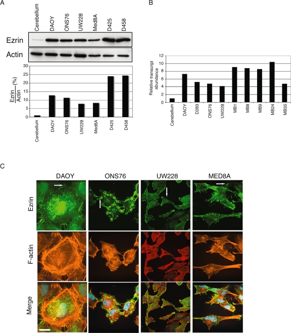
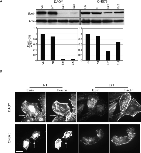
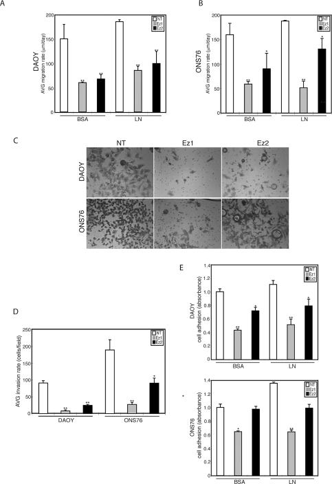
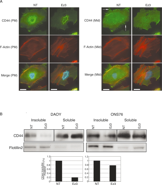
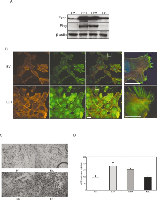
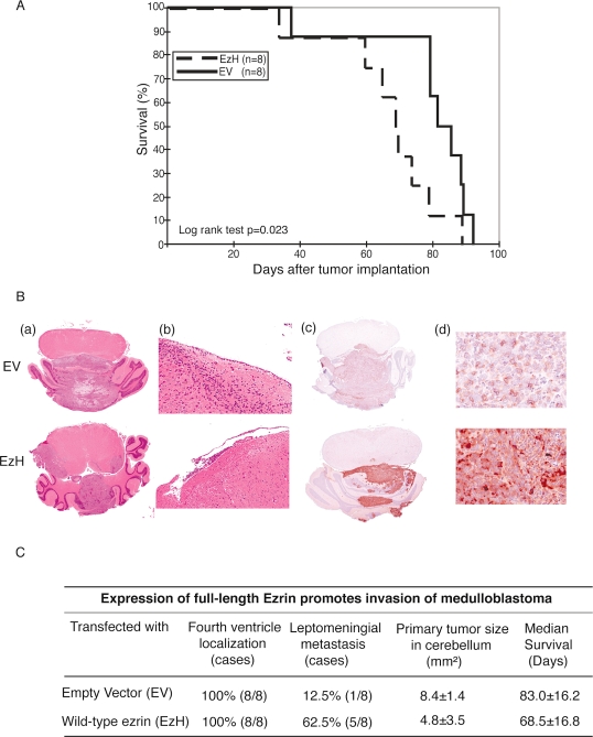
Medulloblastoma is a highly malignant brain tumor that occurs predominantly in children. The molecular pathogenesis of medulloblastoma is under investigation. Previously, we used complementary DNA microarray analysis to compare patterns of gene expression in medulloblastoma samples versus normal cerebellum. The cytoskeletal protein ezrin was found to be overexpressed in medulloblastoma compared with normal cerebellum, an observation that was further validated by immunohistochemistry and real-time PCR analysis. To assess the role of ezrin in medulloblastoma, we studied ezrin's role in medulloblastoma migration, invasion, and adhesion. Western blotting and immunofluorescence showed high expression of ezrin in four medulloblastoma cell lines, and ezrin was primarily localized to filopodia. Ezrin-specific small interfering RNA suppressed the formation of filopodia and in vitro migration, invasion, and adhesion. We also used a stably transfected medulloblastoma cell line to study the effect of ezrin overexpression. We showed that high expression of ezrin promotes filopodia formation and in vitro invasion. Finally, athymic mice implanted with ezrin-overexpressing DAOY medullo-blastoma cell clones in the cerebellum showed shortened survival compared with controls. These findings suggest that, in addition to other cytoskeletal proteins, ezrin plays an important role in medulloblastoma adhesion, migration, and invasion.
Figures
Similar articles
-
Inhibition of tumor cell migration and invasion through knockdown of Rac1 expression in medulloblastoma cells.Cell Mol Neurobiol. 2011 Mar;31(2):251-7. doi: 10.1007/s10571-010-9615-8. Epub 2010 Nov 13. Cell Mol Neurobiol. 2011. PMID: 21076938 Free PMC article.
-
Overexpression of HMGA1 deregulates tumor growth via cdc25A and alters migration/invasion through a cdc25A-independent pathway in medulloblastoma.Acta Neuropathol. 2012 Apr;123(4):553-71. doi: 10.1007/s00401-011-0934-8. Epub 2012 Jan 17. Acta Neuropathol. 2012. PMID: 22249617
-
EphB2 activity plays a pivotal role in pediatric medulloblastoma cell adhesion and invasion.Neuro Oncol. 2012 Sep;14(9):1125-35. doi: 10.1093/neuonc/nos130. Epub 2012 Jun 21. Neuro Oncol. 2012. PMID: 22723427 Free PMC article.
-
Ez-Metastasizing: The Crucial Roles of Ezrin in Metastasis.Cells. 2023 Jun 14;12(12):1620. doi: 10.3390/cells12121620. Cells. 2023. PMID: 37371090 Free PMC article. Review.
-
Filopodia in cell adhesion, 3D migration and cancer cell invasion.Curr Opin Cell Biol. 2015 Oct;36:23-31. doi: 10.1016/j.ceb.2015.06.007. Epub 2015 Jul 14. Curr Opin Cell Biol. 2015. PMID: 26186729 Review.
Cited by
-
FoxM1 influences mouse hepatocellular carcinoma metastasis in vitro.Int J Clin Exp Pathol. 2015 Mar 1;8(3):2771-8. eCollection 2015. Int J Clin Exp Pathol. 2015. PMID: 26045783 Free PMC article.
-
Ezrin phosphorylation on tyrosine 477 regulates invasion and metastasis of breast cancer cells.BMC Cancer. 2012 Mar 7;12:82. doi: 10.1186/1471-2407-12-82. BMC Cancer. 2012. PMID: 22397367 Free PMC article.
-
Regulation of intestinal epithelial cell cytoskeletal remodeling by cellular immunity following gut infection.Mucosal Immunol. 2013 Mar;6(2):369-78. doi: 10.1038/mi.2012.80. Epub 2012 Aug 22. Mucosal Immunol. 2013. PMID: 22910215 Free PMC article.
-
A novel role for ezrin in breast cancer angio/lymphangiogenesis.Breast Cancer Res. 2014 Sep 18;16(5):438. doi: 10.1186/s13058-014-0438-2. Breast Cancer Res. 2014. PMID: 25231728 Free PMC article.
-
A case of early extraneural medulloblastoma metastases in a young adult.Asian J Neurosurg. 2015 Oct-Dec;10(4):331-3. doi: 10.4103/1793-5482.162723. Asian J Neurosurg. 2015. PMID: 26425169 Free PMC article.
References
-
- Louis DN, Pomeroy SL, Cairncross JG. Focus on central nervous system neoplasia. Cancer Cell. 2002;1:125–128. - PubMed
-
- Tarbell NJ, Loeffler JS, Silver B, et al. The change in patterns of relapse in medulloblastoma. Cancer. 1991;68:1600–1604. - PubMed
-
- Rutka JT. Medulloblastoma. Clin Neurosurg. 1997;44:571–585. - PubMed
-
- David KM, Casey AT, Hayward RD, Harkness WF, Phipps K, Wade AM. Medulloblastoma: Is the 5-year survival rate improving? A review of 80 cases from a single institution. J Neurosurg. 1997;86:13–21. - PubMed
-
- Buhren J, Christoph AH, Buslei R, Albrecht S, Wiestler OD, Pietsch T. Expression of the neurotrophin receptor p75NTR in medulloblastomas is correlated with distinct histological and clinical features: Evidence for a medulloblastoma subtype derived from the external granule cell layer. J Neuropathol Exp Neurol. 2000;59:229–240. - PubMed
Publication types
MeSH terms
Substances
LinkOut - more resources
Full Text Sources
