. 2008 Dec 30;105(52):20852-7.
doi: 10.1073/pnas.0810958105. Epub 2008 Dec 16.

Oncogenic kinase NPM/ALK induces through STAT3 expression of immunosuppressive protein CD274 (PD-L1, B7-H1)

Affiliations

Oncogenic kinase NPM/ALK induces through STAT3 expression of immunosuppressive protein CD274 (PD-L1, B7-H1)

Michal Marzec et al. Proc Natl Acad Sci U S A. .

Abstract

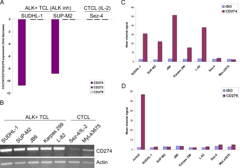
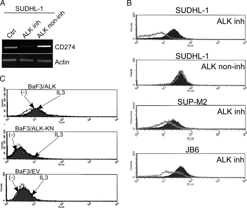
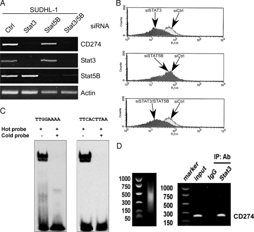
The mechanisms of malignant cell transformation caused by the oncogenic, chimeric nucleophosmin (NPM)/anaplastic lymphoma kinase (ALK) remain only partially understood, with most of the previous studies focusing mainly on the impact of NPM/ALK on cell survival and proliferation. Here we report that the NPM/ALK-carrying T cell lymphoma (ALK+TCL) cells strongly express the immunosuppressive cell-surface protein CD274 (PD-L1, B7-H1), as determined on the mRNA and protein level. The CD274 expression is strictly dependent on the expression and enzymatic activity of NPM/ALK, as demonstrated by inhibition of the NPM/ALK function in ALK+TCL cells by the small molecule ALK inhibitor CEP-14083 and by documenting CD274 expression in IL-3-depleted BaF3 cells transfected with the wild-type NPM/ALK, but not the kinase-inactive NPM/ALK K210R mutant or empty vector alone. NPM/ALK induces CD274 expression by activating its key signal transmitter, transcription factor STAT3. STAT3 binds to the CD274 gene promoter in vitro and in vivo, as shown in the gel electromobility shift and chromatin immunoprecipitation assays, and is required for the PD-L1 gene expression, as demonstrated by siRNA-mediated STAT3 depletion. These findings identify an additional cell-transforming property of NPM/ALK and describe a direct link between an oncoprotein and an immunosuppressive cell-surface protein. These results also provide an additional rationale to therapeutically target NPM/ALK and STAT3 in ALK+TCL. Finally, they suggest that future immunotherapeutic protocols for this type of lymphoma may need to include the inhibition of NPM/ALK and STAT3 to achieve optimal clinical efficacy.


Conflict of interest statement

The authors declare no conflict of interest.

Figures

Fig. 1.
Fig. 1.
CD274 expression by ALK+TCL cells. (A) Expression of CD274 (PD-L1) and the functionally-related CD273 (PD-L2) and CD279 (PD-1) as determined by cDNA oligonucleotide hybridization in ALK+TCL cell lines (SUDHL-1 and SUP-M2), exposed for 6 h to 175 nM of an ALK inhibitor, CEP-14083 or the compound's vehicle. IL-2-dependent, CTCL-derived Sez-4 cell line depleted of IL-2 for 16 h and subsequently exposed for 4 h to IL-2 or medium (20), served as an additional control. The results are depicted as a fold change in the hybridization signal upon cell treatment with the ALK inhibitor or IL-2, as compared to the untreated cells. (B) Expression of CD274 mRNA in the depicted five ALK+TCL and two CTCL cell lines detected by RT-PCR. Expression of actin served as a positive control. Expression of CD274 protein (C) and CD279 (D) at the cell surface of the ALK+TCL and CTCL cell lines detected by flow cytometry. Staining with an isotype-matched antibody of unrelated specificity served as a negative control. Jurkat cell line served as a positive control of CD279 expression.
Fig. 2.
Fig. 2.
Expression of CD274 in ALK+TCL tissues. Section of lymph nodes were examined microscopically using an intermediate (100×, large images) and high (500 and 400×, insets) power magnification. (A) H&E showed a predominance of large, frequently highly atypical cells. Immunohistochemical examination revealed strong, selective staining of the atypical cells by both anti-ALK (B) and anti-CD274 antibodies (C). The depicted images are representative for the 18 ALK+TCL cases examined.
Fig. 3.
Fig. 3.
Expression of CD274 is induced by NPM/ALK. (A) Expression of CD274 mRNA in the ALK+TCL SUDHL-1 cell line before and after treatment with 175 nM of the ALK inhibitor CEP-14083 or its structural analog, CEP-11988, noninhibitory for ALK (ALK non-inh) (14). (B) Expression of CD274 protein in the depicted ALK+TCL cell lines before and after treatment with the ALK inhibitor CEP-14083. Treatment of the SUDHL-1 cell line with the ALK noninhibitory analog CEP-11988 served as a control (lane two). (C) Expression of CD274 examined by flow cytometry in the IL-3-dependent BaF3 cells transfected with the intact, enzymatically active NPM/ALK, kinase-activity negative K210R NPM/ALK mutant (ALK-KN), or empty vector after culture for 48 h in medium with IL-3 or without IL-3 (-).
Fig. 4.
Fig. 4.
NPM/ALK induces CD274 expression through STAT3. (A) The effect of siRNA-mediated STAT3 and STAT5B depletion on CD274 mRNA expression. The ALK+TCL cell line SUDHL-1 was treated with siRNA specific for STAT3 or STAT5, STAT3/STAT5 siRNA combination, or control nonspecific siRNA and evaluated by RT-PCR for expression of mRNA coding for CD274 and the depicted other molecules serving as controls. (B) The effect of the siRNA-mediated STAT3 depletion on expression of the CD274 protein. SUDHL-1 cells treated with the STAT3, STAT5, STAT3/STAT5, or control siRNA were examined for CD274 protein expression by flow cytometry. (C) Binding of STAT3 to the CD274 gene promoter in vitro. The nuclear protein extracts from SUDHL-1 cells were incubated with the hot, biotin-labeled oligonucleotide probes corresponding to either of the two STAT3 binding sites identified within the CD274 gene promoter and analyzed in EMSA. The extract of the SUDHL-1 cells preincubated with the corresponding unlabeled cold probes served as control. (D) Binding of STAT3 to the CD274 gene promoter in vivo. Protein cell lysates from the SUDHL-1 cell line were analyzed in the ChIP assay using an anti-STAT3 rabbit polyclonal antibody and primer pairs specific for CD274 gene promoter. Non-immunoprecipitated lysates (input) and immunoprecipitates obtained with the STAT3 nonimmune entire IgG rabbit serum fraction served as a positive and negative control, respectively.

References

    1. Li R, Morris SW. Development of anaplastic lymphoma kinase (ALK) small-molecule inhibitors for cancer therapy. Med Res Rev. 2008;28:372–412. - PubMed
    1. Chiarle R, Voena C, Ambrogio C, Piva R, Inghirami G. The anaplastic lymphoma kinase in the pathogenesis of cancer. Nat Rev Cancer. 2008;8:11–23. - PubMed
    1. Morris SW, et al. Fusion of a kinase gene, ALK, to a nucleolar protein gene, NPM, in non-Hodgkin's lymphoma. Science. 1994;263:1281–1284. - PubMed
    1. Shiota M, et al. Hyperphosphorylation of a novel 80 kDa protein-tyrosine kinase similar to Ltk in a human Ki-1 lymphoma cell line, S3. Oncogene. 1994;9:1567–1574. - PubMed
    1. Morris SW, et al. ALK, the chromosome 2 gene locus altered by the t (2; 5) in non-Hodgkin's lymphoma, encodes a novel neural receptor tyrosine kinase that is highly related to leukocyte tyrosine kinase (LTK) Oncogene. 1997;14:2175–2188. - PubMed

Publication types

MeSH terms