

doi: 10.1099/vir.0.006353-0.
Upstream-binding factor is sequestered into herpes simplex virus type 1 replication compartments
Affiliations
- PMID: 19088274
- PMCID: PMC2885023
- DOI: 10.1099/vir.0.006353-0
Item in Clipboard
Upstream-binding factor is sequestered into herpes simplex virus type 1 replication compartments
J Gen Virol.
2009 Jan.
Abstract
Previous reports have shown that adenovirus recruits nucleolar protein upstream-binding factor (UBF) into adenovirus DNA replication centres. Here, we report that despite having a different mode of viral DNA replication, herpes simplex virus type 1 (HSV-1) also recruits UBF into viral DNA replication centres. Moreover, as with adenovirus, enhanced green fluorescent protein-tagged fusion proteins of UBF inhibit viral DNA replication. We propose that UBF is recruited to the replication compartments to aid replication of HSV-1 DNA. In addition, this is a further example of the role of nucleolar components in viral life cycles.
Figures
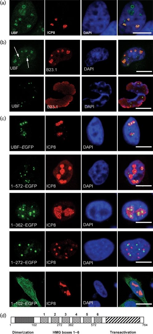
UBF is enriched in viral replication centres. (a) Cells infected with HSV-1 and probed with ICP8 antibodies at 7 h p.i. The location of UBF is shown in green, ICP8 protein is in red and the extent of the cell nucleus is revealed by staining with DAPI in blue. (b) Infected cells at 7 h p.i. (top row) and 24 h p.i. (bottom row). Arrows point to sites of extra nucleolar accumulation of UBF. The location of UBF is in green, B23.1 is in red and DAPI is in blue. All images are of a single focal plane approximately 0.3 μm in depth. Bar, 10 μm. (c) Location of a number of UBF–EGFP fusion proteins in HeLa cells at 7 h p.i. In each case, the plasmid indicated had been transfected into the cells approximately 15 h prior to infection. The EGFP fusion is shown in green, ICP8 is in red and DAPI is in blue. UBF–EGFP contains the full-length protein. (d) Schematic diagram of UBF. The drawing indicates the relative location of the 6 HMG boxes (numbered 1–6), the dimerization domain (marked with checked fill) and the transactivation domain (marked with diagonal lines). In addition, below the diagram the approximate locations of the C-terminal amino acids of the deletion mutants used in this study (full-length UBF is 764 aa) are indicated.
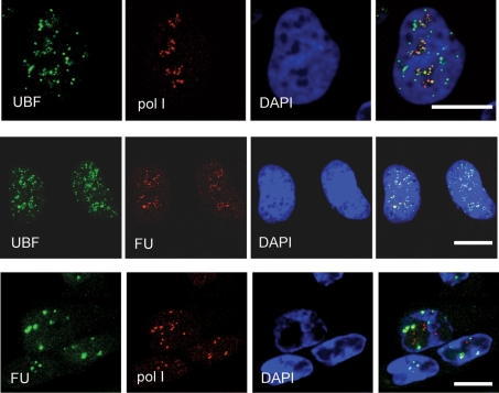
HSV-1 infection causes UBF to separate from RNA pol I and the major sites of RNA synthesis. HSV-1-infected cells at 7 h p.i., where the top row shows endogenous UBF (green), RNA pol I (red) and DAPI (blue). Similarly infected cells are shown in the middle row, endogenous UBF (green), RNA synthesis (detected by FU labelling for 20 min; red) and DAPI (blue) are shown. Similarly infected cells are also shown in the bottom row, where the location of FU (green), RNA pol I (red) and DAPI (blue) are shown. All images are of a single focal plane approximately 0.3 μm in depth. Bar, 10 μm.

The effects of HSV-1 infection on UBF and the effects of UBF fusion proteins on HSV-1 replication. In (a) the levels of total UBF and phosporylated UBF were assessed by Western blotting. On the left is a Western analysis of cells harvested at 7 and 24 h p.i., showing levels of total UBF (both isoforms) by using an affinity purified anti-UBF serum raised in sheep. The same samples were analysed for phosphorylated ser 388 UBF. Also shown is a control Western blot using antisera to actin as a loading control. (b) Shows the effect of expression of UBF–EGFP fusion proteins on HSV-1 origin-dependent DNA synthesis in the presence of the indicated UBF fusion proteins or pE9CT. The position of the replicated pS1 molecules are indicated by the arrow. (c) Shows the effects of the indicated plasmids on the ability of infectious HSV-1 DNA to generate plaques in a co-transfection assay.
Similar articles
-
Upstream binding factor inhibits herpes simplex virus replication.Virology. 2015 Sep;483:108-16. doi: 10.1016/j.virol.2015.04.003. Epub 2015 May 15. Virology. 2015. PMID: 25965800
-
Relocalization of upstream binding factor to viral replication compartments is UL24 independent and follows the onset of herpes simplex virus 1 DNA synthesis.J Virol. 2010 May;84(9):4810-5. doi: 10.1128/JVI.02437-09. Epub 2010 Feb 10. J Virol. 2010. PMID: 20147409 Free PMC article.
-
Nucleolar protein upstream binding factor is sequestered into adenovirus DNA replication centres during infection without affecting RNA polymerase I location or ablating rRNA synthesis.J Cell Sci. 2006 Jun 15;119(Pt 12):2621-31. doi: 10.1242/jcs.02982. J Cell Sci. 2006. PMID: 16763197
-
HSV-1 DNA Replication-Coordinated Regulation by Viral and Cellular Factors.Viruses. 2021 Oct 7;13(10):2015. doi: 10.3390/v13102015. Viruses. 2021. PMID: 34696446 Free PMC article. Review.
-
Molecular mechanisms of replication of herpes simplex virus 1.Acta Virol. 2000 Oct;44(5):289-307. Acta Virol. 2000. PMID: 11252674 Review.
Cited by
-
Nucleolar proteomics and viral infection.Proteomics. 2010 Nov;10(22):4077-86. doi: 10.1002/pmic.201000251. Proteomics. 2010. PMID: 20661956 Free PMC article. Review.
-
Involvement of the nucleolus in replication of human viruses.Rev Med Virol. 2009 Jul;19(4):201-14. doi: 10.1002/rmv.614. Rev Med Virol. 2009. PMID: 19399920 Free PMC article. Review.
-
Viruses and the nucleolus: the fatal attraction.Biochim Biophys Acta. 2014 Jun;1842(6):840-7. doi: 10.1016/j.bbadis.2013.12.010. Epub 2013 Dec 27. Biochim Biophys Acta. 2014. PMID: 24378568 Free PMC article.
-
HIV-1 infection causes a down-regulation of genes involved in ribosome biogenesis.PLoS One. 2014 Dec 2;9(12):e113908. doi: 10.1371/journal.pone.0113908. eCollection 2014. PLoS One. 2014. PMID: 25462981 Free PMC article.
-
Evidence for ubiquitin-regulated nuclear and subnuclear trafficking among Paramyxovirinae matrix proteins.PLoS Pathog. 2015 Mar 17;11(3):e1004739. doi: 10.1371/journal.ppat.1004739. eCollection 2015 Mar. PLoS Pathog. 2015. PMID: 25782006 Free PMC article.
References
-
- Bertrand, L. & Pearson, A. (2008). The conserved N-terminal domain of herpes simplex virus 1 UL24 protein is sufficient to induce the spatial redistribution of nucleolin. J Gen Virol 89, 1142–1151. - PubMed
-
- Besse, S. & Puvion-Dutilleul, F. (1996). Intranuclear retention of ribosomal RNAs in response to herpes simplex virus type 1 infection. J Cell Sci 109, 119–129. - PubMed
-
- Boehmer, P. E. & Lehman, I. R. (1997). Herpes simplex virus DNA replication. Annu Rev Biochem 66, 347–384. - PubMed
-
- Boisvert, F. M., van Koningsbruggen, S., Navascues, J. & Lamond, A. I. (2007). The multifunctional nucleolus. Nat Rev Mol Cell Biol 8, 574–585. - PubMed
Publication types
MeSH terms
Substances
Grants and funding
LinkOut - more resources
Full Text Sources