. 2009 Jan;90(Pt 1):177-86.
doi: 10.1099/vir.0.005678-0.

Inhibition of mRNA export and dimerization of interferon regulatory factor 3 by Theiler's virus leader protein

Affiliations

Inhibition of mRNA export and dimerization of interferon regulatory factor 3 by Theiler's virus leader protein

Céline Ricour et al. J Gen Virol. 2009 Jan.

Abstract

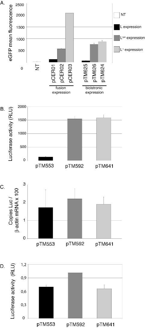
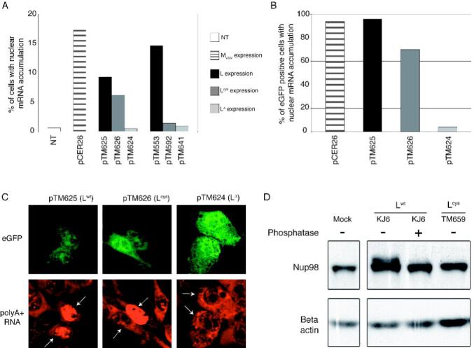
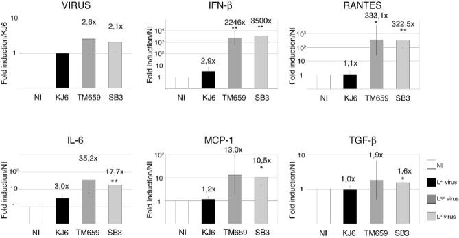
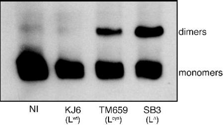
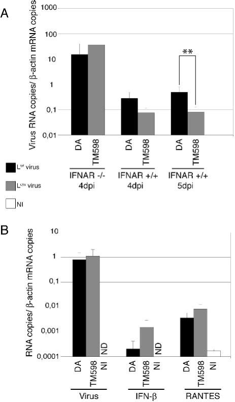
Theiler's murine encephalomyelitis virus (TMEV or Theiler's virus) is a neurotropic picornavirus that can persist lifelong in the central nervous system of infected mice, causing a chronic inflammatory demyelinating disease. The leader (L) protein of the virus is an important determinant of viral persistence and has been shown to inhibit transcription of type I interferon (IFN) genes and to cause nucleocytoplasmic redistribution of host proteins. In this study, it was shown that expression of the L protein shuts off synthesis of the reporter proteins green fluorescent protein and firefly luciferase, suggesting that it induces a global shut-off of host protein expression. The L protein did not inhibit transcription or translation of the reporter genes, but blocked cellular mRNA export from the nucleus. This activity correlated with the phosphorylation of nucleoporin 98 (Nup98), an essential component of the nuclear pore complex. In contrast, the data confirmed that the L protein inhibited IFN expression at the transcriptional level, and showed that transcription of other chemokine or cytokine genes was affected by the L protein. This transcriptional inhibition correlated with inhibition of interferon regulatory factor 3 (IRF-3) dimerization. Whether inhibition of IRF-3 dimerization and dysfunction of the nuclear pore complex are related phenomena remains an open question. In vivo, IFN antagonism appears to be an important role of the L protein early in infection, as a virus bearing a mutation in the zinc finger of the L protein replicated as efficiently as the wild-type virus in type I IFN receptor-deficient mice, but had impaired fitness in IFN-competent mice.


Figures

Figure 1
Figure 1. Post-transcriptional shut-off of cellular protein synthesis by the TMEV leader protein
BALB/3T3 cells were transfected with constructs encoding L, Lcys and LΔ. Histograms show the means and Standard Deviation of one representative experiment performed in triplicate. A. eGFP mean fluorescence measured by FACS, 24 hours after transfection of the plasmids encoding the L-eGFP fusions or the L-IRES-eGFP constructs. (NT: non transfected cells) B. Luciferase activity (relative luciferase units) measured 7 hours after co-transfection of cells with a plasmid expressing luciferase (pCS41) and either L (pTM553), Lcys (pTM592) or LΔ6-67 (pTM641). C. Amount of luciferase mRNA, measured by real-time RT-PCR in cells co-transfected as described in B. Values were normalized to β-actin. Note that no effect of L on β-actin mRNA levels was detected at that time point. D. Leader does not affect translation of cytoplasmic mRNA. BALB/3T3 cells were co-transfected with luciferase capped RNA and with plasmid DNA (ratio 1/10) coding for L (pTM533), Lcys (pTM592) and LΔ6-67 (pTM641). Histograms show luciferase activity, measured 14 hours after co-transfection.
Figure 2
Figure 2. The leader promotes nuclear retention of cellular polyA+ RNA and triggers phosphorylation of Nup98
A. BALB/3T3 cells were transfected with L, Lcys or LΔ6-67 expression plasmids (pTM553, pTM592 and pTM641, respectively) or with the bicistronic constructs co-expressing these proteins and eGFP (pTM625, pTM626 and pTM624, respectively). The bicistronic construct expressing VSV M protein (pCER26) was used as control. PolyA+ RNA was detected by fluorescent in situ hybridization using an oligo(dT) probe. The percentage of cells showing higher fluorescent intensity in the nucleus than in the cytoplasm at 16 hours after transfection is shown. (NT: non transfected cells). B. Percentage of cells showing higher fluorescent intensity in the nucleus, among 50 eGFP positive cells. C. Representative ISH results in cells transfected with the bicistronic constructs, showing eGFP fluorescence (above) and detection of polyA+ RNA by ISH (below). In the latter panel, white arrows point to eGFP positive cells. D. Phosphorylation of Nup98 in cells infected with TMEV. BHK-21 cells were infected for 12 hours with either KJ6 or TM659 or were mock-infected. Lysates were prepared and analyzed by SDS PAGE and immunoblotting to detect Nup98. Phosphatase indicates whether protein extracts were treated with calf alkaline phosphatase.
Figure 3
Figure 3. Influence of the TMEV leader on the transcription of cytokine and chemokine genes
A. L929 cells were infected for 9 hours with 2 PFU per cell of either KJ6 (Lwt) (black), TM659 (Lcys) (medium grey), or SB3 (LΔ6-67) (light grey), or were mock-infected (NI) (white). mRNA levels, detected by comparative real-time RT-PCR, were normalized to the amounts of β-actin for each sample. Histograms show the means and SD of relative gene expressions. Ratios to KJ6 infected samples are shown in the case of viral RNA. Ratios to mock-infected cells are shown for other genes. * and ** indicate significant differences with the corresponding KJ6-infected sample (p values < 0.05 and <0.01 respectively, determined in an unpaired t-test).
Figure 4
Figure 4. Inhibition of IRF-3 dimerization
L929 cells were infected for 9 hours with either KJ6 (L), TM659 (Lcys), or SB3 (LΔ6-67), or were mock-infected (NI). Protein extracts were separated by acrylamide gel electrophoresis under non-denaturing conditions and IRF-3 dimer formation was detected by immunoblotting.
Figure 5
Figure 5. Comparison of viral load and IFN-β and RANTES gene expression in brains of infected mice
Real-time quantitative PCR was used to measure viral RNA or IFN-β and RANTES mRNA in total RNA extracted from the brain of infected mice. A. Comparison of Lwt (DA1) and Lcys (TM598) viral RNA amounts (mean and SD) in the brains of IFNAR -/- or IFNAR +/+ mice, 4 and 5 days post-infection. B. Viral genomes, RANTES mRNA and IFN-β mRNA levels measured in the brain of IFNAR -/- mice infected with Lwt (DA1) and Lcys (TM598) viruses, or mock-infected (NI), 1 day post-inoculation. ND indicates that the cDNA copy number was under the detection threshold (<10-4 copies per copy of β-actin cDNA). **: t test, p < 0.01.

Similar articles

Cited by

References

    1. Baida G, Popko B, Wollmann RL, Stavrou S, Lin W, Tretiakova M, Krausz TN, Roos RP. A subgenomic segment of Theiler’s murine encephalomyelitis virus RNA causes demyelination. J Virol. 2008;82:5879–5886. - PMC - PubMed
    1. Blevins MB, Smith AM, Phillips EM, Powers MA. Complex formation among the RNA export proteins Nup98, Rae1/Gle2, and TAP. J Biol Chem. 2003;278:20979–20988. - PubMed
    1. Brahic M, Bureau JF, Michiels T. The Genetics of the Persistent Infection and Demyelinating Disease Caused by Theiler’s Virus. Annu Rev Microbiol. 2005 - PubMed
    1. Chakraborty P, Satterly N, Fontoura BM. Nuclear export assays for poly(A) RNAs. Methods. 2006;39:363–369. - PubMed
    1. Chen HH, Kong WP, Roos RP. The leader peptide of Theiler’s murine encephalomyelitis virus is a zinc-binding protein. J Virol. 1995;69:8076–8078. - PMC - PubMed

Publication types

MeSH terms

LinkOut - more resources