. 2008 Dec;20(12):3374-88.
doi: 10.1105/tpc.108.063859. Epub 2008 Dec 16.

Arabidopsis synaptotagmin 1 is required for the maintenance of plasma membrane integrity and cell viability

Affiliations

Arabidopsis synaptotagmin 1 is required for the maintenance of plasma membrane integrity and cell viability

Arnaldo L Schapire et al. Plant Cell. 2008 Dec.

Abstract

Plasma membrane repair in animal cells uses synaptotagmin 7, a Ca(2+)-activated membrane fusion protein that mediates delivery of intracellular membranes to wound sites by a mechanism resembling neuronal Ca(2+)-regulated exocytosis. Here, we show that loss of function of the homologous Arabidopsis thaliana Synaptotagmin 1 protein (SYT1) reduces the viability of cells as a consequence of a decrease in the integrity of the plasma membrane. This reduced integrity is enhanced in the syt1-2 null mutant in conditions of osmotic stress likely caused by a defective plasma membrane repair. Consistent with a role in plasma membrane repair, SYT1 is ubiquitously expressed, is located at the plasma membrane, and shares all domains characteristic of animal synaptotagmins (i.e., an N terminus-transmembrane domain and a cytoplasmic region containing two C2 domains with phospholipid binding activities). Our analyses support that membrane trafficking mediated by SYT1 is important for plasma membrane integrity and plant fitness.


Figures

Figure 1.
Figure 1.
Identification of Mutants in the SYT1 Gene and Expression Analyses. (A) Wild-type C-24 and syt1-2 seedlings were grown for 3 to 4 d in half-strength Murashige and Skoog (MS) medium (1.5 mM Ca2+) and then transferred to the same medium (left panel) and medium containing 160 mM NaCl (right panel) and photographed 20 d later. (B) T-DNA insertion sites in syt1-2 and syt1-2 with exons shown as gray boxes. (C) Diagram of the structure of the animal synaptotagmins (top) and Arabidopsis SYT1 (bottom). SYT1 is predicted to have a short extracellular domain (blue), a transmembrane domain (gray), a linker domain (yellow), and two tandem intracellular C2 domains, C2A and C2B. (D) RT-PCR analysis using primers flanking the T-DNA insertion in the syt1-2 mutant (top). Immunoblots probed with antisera to SYT1 peptides indicate that no SYT1 protein is produced in syt1-2 (bottom). The molecular weight indicated was calculated from the protein gel blot. (E) RT-PCR analysis of SYT1 in root (1), shoot (2), rosette leaves (3), cauline leaves (4), inflorescence (5), and silique (6). A genomic control was used (7). (F) SYT1 promoter-driven GUS expression in wild-type seedlings is observed in all tissues analyzed. (a) A 3-d-old seedling root; (b) a 2-week-old plant; (c) trichomes; (d) root of a 2-week-old plant; (e) inflorescence.
Figure 2.
Figure 2.
Subcellular Localization of SYT1. (A) Expression of SYT1:GFP shows stable bright spots (arrows) at the plasma membrane connected by ER tubules (arrowheads). Bar = 25 μm. (B) Leaf cells were plasmolyzed using 500 mM NaCl for 2 h. The middle of the cell is shown in a single optical section. The GFP signal remains in the retracted Hechtian strands at the plasma membrane and at the cell wall (left). FM4-64 staining of the same cell shows the stained plasma membrane and spots in the cell wall (middle). A merge of the two images reveals the colocalization of the SYT1:GFP spots to the plasma membrane and membrane positive spots at the cell wall (right). Bar = 25 μm. (C) Projection of a salt plasmolyzed cell showing SYT1:GFP in Hechtian strands (arrow) and at the shrunken protoplast (arrowheads). Bar = 25 μm. (D) to (F) Transgenic Arabidopsis expressing 35S-SYT1:GFP. (D) Projection of cells of the lower epidermis of leaves. Nonmotile spots residing at the plasma membrane/cell wall are labeled. The inset shows a single optical section from the middle of the cell. Autofluorescence of open stomata cell wall and plastids appear as bright green ring and bright yellow structures, respectively. Bar = 25 μm. (E) Detail view of epidermal cells in the elongation region. Bar = 10 μm. (F) Projection of a root tip. Bar = 50 μm.
Figure 3.
Figure 3.
Preferential Plasma Membrane Localization of SYT1 Protein. (A) to (D) Confocal immunohistochemical images of SYT1 in wild-type plants. (A) Cross section of a root apex. (B) Detailed view of cross-walls in the transition zone of the root apex enriched in SYT1:GFP. Bar = 10 μm. (C) Shoot apex cells. (D) Cotyledon cells. The 4',6-diamidino-2-phenylindole–positive nuclei appear as blue structures. Bars in (A), (C), and (D) = 25 μm. (E) and (F) Immunogold electron microscopy localization confirms the plasma membrane localization of SYT1. (E) Young emerging cell wall (star) with some membraneous structures as traces of the endosomal origin of cytokinetic cell plate (arrow). Note large aggregates of SYT1-positive gold particles near cell plate (arrowhead). (F) Cell wall (star) with young plasmodesmata is decorated with the SYT1-positive gold particles (arrowheads), while more mature plasmodesmata show gold particles near their orifices (arrows). The abundance of the gold particles at the plasma membrane is lower under more mature cell wall. Bars = 500 nm.
Figure 4.
Figure 4.
Ca2+- and Phospholipid-Dependent Membrane Binding by SYT1. (A) Sequence alignment of the C2 domains from rat synaptotagmin 1 (RS1), rat synaptotagmin 7 (RS7), and SYT1. Similar colors indicate similar polarity of the residues. Amino acid residues that form the Ca2+ binding sites of the C2 domains in rat synaptotagmin 1 and 7 are indicated by arrowheads. (B) Recombinant GST fusion proteins containing the residues indicated were used in phospholipid binding assays. Proteins were incubated in the absence (−) or the presence (+) of 100 μM Ca2+ with liposomes composed of 25% PS/75% PC. Liposomes were precipitated by centrifugation, and bound proteins were analyzed by SDS-PAGE. (C) to (E) The C2A domain (C), C2B domain (D), and C2AB domains (E) of SYT1 were incubated with liposomes as in (B) in the presence of the indicated concentrations of free Ca2+ (clamped with Ca2+/EGTA buffers) to determine the half-maximal binding for free Ca2+. The EC50 for SYT1-C2A was 6 ± 0.6 μM (paired t test, P < 0.05; n = 4). No binding was obtained when the liposomes were incubated in the presence of GST alone.
Figure 5.
Figure 5.
Physiological Characterization of the syt1-2 Mutant. (A) Phenotypes of wild-type and syt1-2 seedlings growing at increasing NaCl concentrations. Wild-type and syt1-2 seedlings were grown for 3 d in half-strength MS medium and then transferred to one-tenth MS medium at the Ca2+ and NaCl concentrations indicated and photographed 7 d later. The seedlings shown are representative of four independent trials. Bar = 10 mm; inset bars = 1 mm. (B) Quantification of fresh weight for seedlings treated as described for (A). For fresh weight determination, 12 seedlings were weighed at one time and the result was divided by 12. Data are means ± se (n = 4). All mean values are significantly different between the wild type and syt1-2 (P < 0.05) with the exception of the 200 mM NaCl treatment in which no significant differences were found. (C) One-week-old seedlings grown in half-strength MS medium without NaCl were transferred to one-tenth MS medium plus the indicated NaCl and Ca2+ concentrations, and after 12 h electrolyte leakage was determined. Data are means of three independent assays. All mean values are significantly different between the wild type and syt1-2 (P < 0.05). (D) Ion leakage from 1-week-old seedlings treated with PEG. Data are means of three independent assays. All mean values are significantly different between the wild type and syt1-2 (P < 0.05). (E) Ion leakage from 1-week-old seedlings treated with PEG performed as in (D), but root and shoot tissues were separated after PEG treatment. Data are means of three independent assays. All mean values are significantly different between the wild type and syt1-2 (P < 0.05).
Figure 6.
Figure 6.
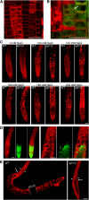
syt1-2 Shows Reduced Cell Survival and Plasma Membrane Viability after NaCl Treatment. (A) Massive staining of Arabidopsis root cells is the result of loss of plasma membrane integrity. A root from a wild-type plant was pressed between glass slides to induce mechanical damage and then stained with FM4-64. Bar = 25 μm. (B) Double staining using FM4-64 and FDA. Viable cells that are stained in green by FDA show intact plasma membrane (arrow). Bar = 25 μm. (C) Plasma membrane integrity analysis of syt1-2 and wild-type roots by FM4-64 staining after 1 h of NaCl treatment. Representative roots from five independent experiments are shown. Bar = 50 μm. (D) Roots of syt1-2 after 1 h in 200 mM NaCl. The red (FM4-64) channel (left), the green (FDA) channel (middle), and the merged image (right) are shown. A closer view is shown in the right panel. Bars = 50 μm. (E) Three-day-old seedlings grown in half-strength MS were transferred to one-tenth MS plates plus 150 mM NaCl. After 1 week of treatment, seedlings were stained with FM4-64. White arrows indicate the position of the root apex at the beginning of NaCl treatment. For illustration purposes, two images were joined (indicated by arrowheads) using Adobe Photoshop. Bar = 100 μm.

Comment in

References

    1. Alexandersson, E., Saalbach, G., Larsson, C., and Kjellbom, P. (2004). Arabidopsis plasma membrane proteomics identifies components of transport, signal transduction and membrane trafficking. Plant Cell Physiol. 45 1543–1556. - PubMed
    1. Andrews, N. (2005). Membrane resealing: Synaptotagmin VII keeps running the show. Sci. STKE 2005 pe19. - PubMed
    1. Andrews, N., and Chakrabarti, S. (2005). There's more to life than neurotransmission: The regulation of exocytosis by synaptotagmin VII. Trends Cell Biol. 15 626–631. - PubMed
    1. Baena-González, E., Rolland, F., Thevelein, J., and Sheen, J. (2007). A central integrator of transcription networks in plant stress and energy signalling. Nature 448 938–942. - PubMed
    1. Baluska, F., Menzel, D., and Barlow, P.W. (2006). Cytokinesis in plant and animal cells: Endosomes 'shut the door'. Dev. Biol. 294 1–10. - PubMed

Publication types

MeSH terms