. 2009 Feb;5(2):217-20.
doi: 10.4161/auto.5.2.7201. Epub 2009 Feb 17.

Indirect estimation of the area density of Atg8 on the phagophore

Affiliations

Indirect estimation of the area density of Atg8 on the phagophore

Zhiping Xie et al. Autophagy. 2009 Feb.

Abstract

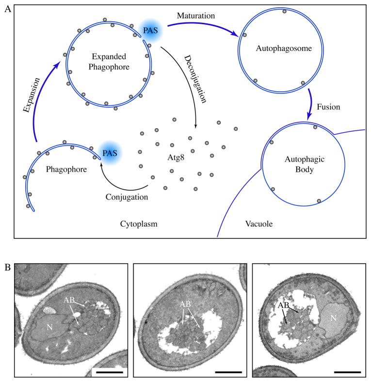
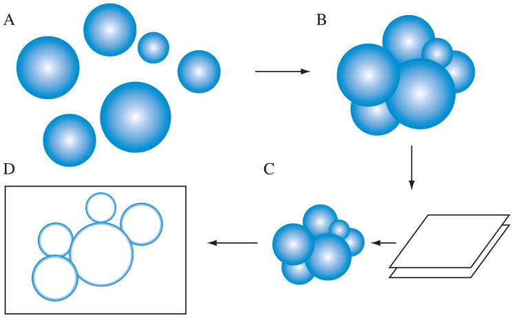
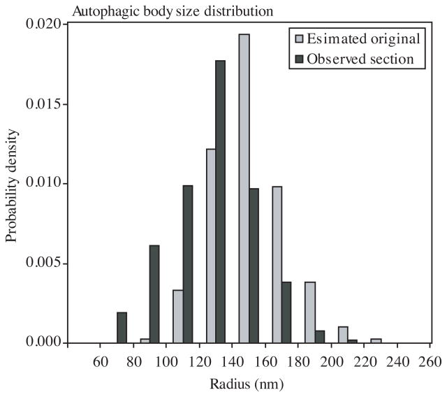
Atg8 is a ubiquitin-like protein that controls the expansion of the phagophore during autophagosome formation. It is recruited to the phagophore during the expansion stage and released upon the completion of the autophagosome. One possible model explaining the function of Atg8 is that it acts as an adaptor of a coat complex. Here, we tested the coat-adaptor model by estimating the area density of Atg8 molecules on the phagophore. We developed a computational process to simulate the random sectioning of vesicles heterogeneous in size. This method can be applied to estimate the original sizes of intracellular vesicles from sizes of their random sections obtained through transmission electron microscopy. Using this method, we found that the estimated area density of Atg8 is comparable with that of proteins that form the COPII coat.


Figures

Figure 1
Figure 1
Formation of autophagosomes and autophagic bodies. (A) Scheme of autophagy in yeast. Autophagosomes are formed through the expansion and deformation of the phagophore at the PAS. During this process, Atg8 is conjugated and recruited to the PAS. It resides on both sides of the phagophore and controls its expansion. At the end of phagophore expansion, most Atg8 molecules are released back to the cytosol through deconjugation. The fully expanded phagophore then matures into an autophagosome. When autophagosomes fuse with the yeast vacuole, the inner vesicles are released into the vacuole lumen, and are now termed autophagic bodies. In pep4Δ cells, autophagic bodies are not degraded. (B) Representative sections of starving pep4Δ yeast cells. atg8Δ pep4Δ cells expressing GFP-Atg8 and grown to mid-log phase were incubated in nitrogen starvation medium for 4 hours and processed for electron microscopy. Autophagic bodies (AB) accumulated as a cluster of single-membrane vesicles in the yeast vacuole. N, nucleus, Scale bar, 1 μm.
Figure 2
Figure 2
Scheme for the computational simulation of sectioning autophagic bodies. (A) First, the program generates a population of vesicles based on the specified parameters. (B) Next, the vesicles are positioned so that their surfaces contact each other. (C) The vesicle cluster is sectioned to produce a 70 nm thick slice. (D) The areas of vesicle sections in the slice is quantified and reported.
Figure 3
Figure 3
The size distributions of observed sections and estimated original vesicles. Observed section, sizes of actually observed sections of autophagic bodies from EM. Estimated original, the sizes of original autophagic bodies estimated from simulation results.

References

    1. Xie Z, Klionsky DJ. Autophagosome formation: core machinery and adaptations. Nat Cell Biol. 2007;9:1102–9. - PubMed
    1. Wang C-W, Stromhaug PE, Shima J, Klionsky DJ. The Ccz1-Mon1 protein complex is required for the late step of multiple vacuole delivery pathways. J Biol Chem. 2002;277:47917–27. - PMC - PubMed
    1. Wang C-W, Stromhaug PE, Kauffman EJ, Weisman LS, Klionsky DJ. Yeast homotypic vacuole fusion requires the Ccz1-Mon1 complex during the tethering/docking stage. J Cell Biol. 2003;163:973–85. - PMC - PubMed
    1. Bonifacino JS, Glick BS. The mechanisms of vesicle budding and fusion. Cell. 2004;116:153–66. - PubMed
    1. Stagg SM, LaPointe P, Balch WE. Structural design of cage and coat scaffolds that direct membrane traffic. Curr Opin Struct Biol. 2007;17:221–8. - PubMed

MeSH terms

Substances