

Histomorphometric analysis of pure titanium implants with porous surface versus rough surface
- PMID: 19089076
- PMCID: PMC4327200
- DOI: 10.1590/s1678-77572006000300013
Histomorphometric analysis of pure titanium implants with porous surface versus rough surface
Abstract
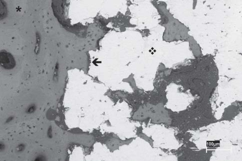
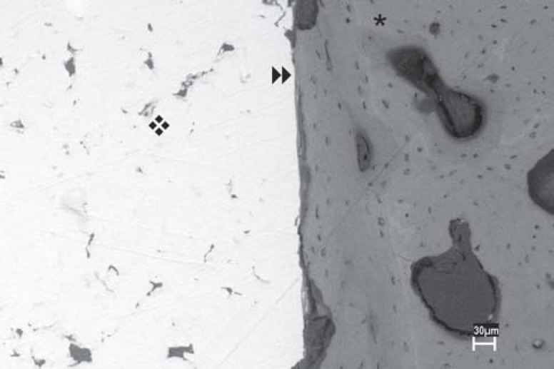
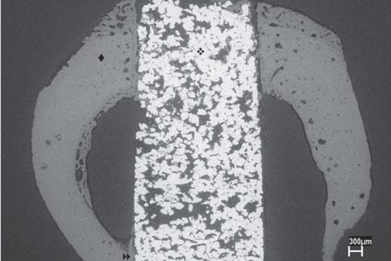
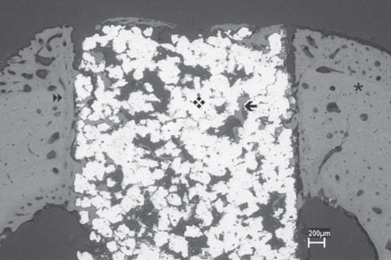
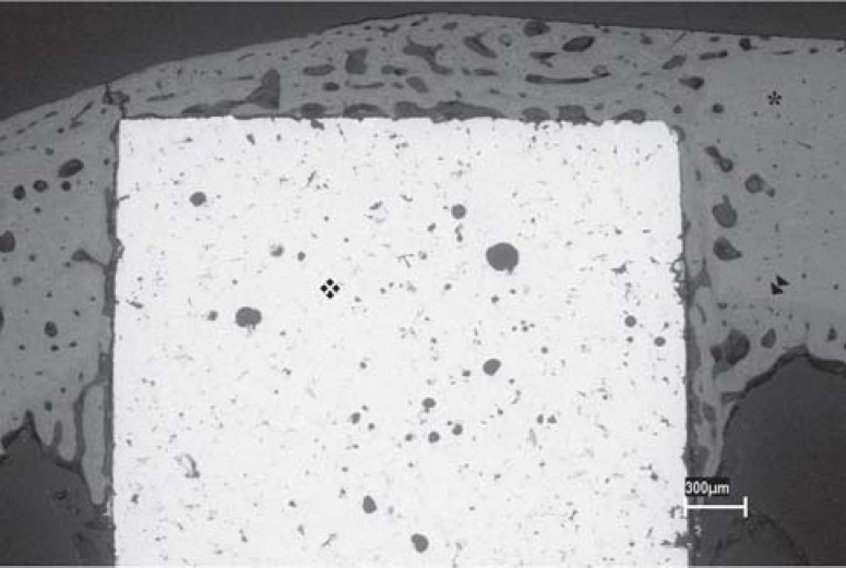
The purpose of this study was to analyze the bone repair around commercially pure titanium implants with rough and porous surface, fabricated using powder metallurgy technique, after their insertion in tibiae of rabbits. Seven male rabbits were used. Each animal received 3 porous-surface implants in the left tibia and 3 rough-surface implants in the right tibia. The rabbits were sacrificed 4 weeks after surgery and fragments of the tibiae containing the implants were submitted to histological and histomorphometric analyses to evaluate new bone formation at the implant-bone interface. Means (%) of bone neoformation obtained in the histomorphometric analysis were compared by Student's t-test for paired samples at 5% significance level.. The results of the histological analysis showed that osseointegration occurred for both types of implants with similar quality of bone tissue. The histomorphometric analysis revealed means of new bone formation at implant-bone interface of 79.69 +/- 1.00% and 65.05 +/- 1.23% for the porous- and rough-surface implants, respectively. Statistically significant difference was observed between the two types of implants with respect to the amount new bone formation (p<0.05). In conclusion, the porous-surface implants contributed to the osseointegration because they provide a larger contact area at implant-bone interface.
Opropósito deste estudo foi avaliar a reparação óssea ao redor de implantes de superfície porosa comparados com implantes de superfície rugosa, ambos confeccionados de titânio puro grau 2 por meio da técnica de metalurgia do pó. Os implantes foram inseridos em tíbias de coelhos. Foram utilizados neste estudo 7 coelhos machos, sendo que cada um recebeu 3 implantes de superfície porosa na tíbia esquerda e 3 implantes de superfície rugosa na tíbia direita. Os animais foram sacrificados 4 semanas após a cirurgia e os fragmentos das tíbias contendo os implantes foram submetidos à análise histológica e histomorfométrica, visando analisar a neoformação óssea na interface osso-implante. As médias (%) obtidas na análise histomorfométrica foram avaliadas por meio do teste estatístico t-student de amostras pareadas com nível de significância de 5%. Os resultados da análise histológica mostraram que a osseointegração foi obtida nos dois tipos de implantes com similar qualidade de tecido ósseo. Na análise histomorfométrica, verificaram-se médias de neoformação óssea na interface osso-implante de 79,69% ± 1,00 e 65,05 ± 1,23 para os implantes de superfície porosa e rugosa, respectivamente, e foi observada diferença estatisticamente significante entre os dois tipos de implantes com relação à quantidade de neoformação óssea. Concluiu-se que os implantes de superfície porosa contribuíram para a osseointegração devido à sua maior superfície de contato na interface osso-implante.
Figures
Similar articles
-
Evaluation of bone ingrowth into porous titanium implant: histomorphometric analysis in rabbits.Braz Oral Res. 2010 Oct-Dec;24(4):399-405. doi: 10.1590/s1806-83242010000400005. Braz Oral Res. 2010. PMID: 21180959
-
In vivo osteogenesis and in vitro Streptococcus mutans adherence: porous-surfaced cylindrical implants vs rough-surfaced threaded implants.Int J Oral Maxillofac Implants. 2013 Nov-Dec;28(6):1630-8. doi: 10.11607/jomi.2747. Int J Oral Maxillofac Implants. 2013. PMID: 24278932
-
Effects on the osseointegration of titanium implants incorporating calcium-magnesium: a resonance frequency and histomorphometric analysis in rabbit tibia.Clin Oral Implants Res. 2018 Jul;29(7):785-791. doi: 10.1111/clr.12909. Epub 2016 Jul 6. Clin Oral Implants Res. 2018. PMID: 27381553
-
Bone response to a pure titanium implant surface modified by laser etching and microarc oxidation.Int J Oral Maxillofac Implants. 2010 Jan-Feb;25(1):130-6. Int J Oral Maxillofac Implants. 2010. PMID: 20209195
-
Osseointegration of titanium, titanium alloy and zirconia dental implants: current knowledge and open questions.Periodontol 2000. 2017 Feb;73(1):22-40. doi: 10.1111/prd.12179. Periodontol 2000. 2017. PMID: 28000277 Review.
Cited by
-
Sequential osseointegration from osseohealing to osseoremodeling - Histomorphological comparison of novel 3D porous and solid Ti-6Al-4V titanium implants.Histol Histopathol. 2021 Jul;36(7):753-764. doi: 10.14670/HH-18-333. Epub 2021 Mar 29. Histol Histopathol. 2021. PMID: 33779981
-
Mechanical Characterisation and Numerical Modelling of TPMS-Based Gyroid and Diamond Ti6Al4V Scaffolds for Bone Implants: An Integrated Approach for Translational Consideration.Bioengineering (Basel). 2022 Sep 24;9(10):504. doi: 10.3390/bioengineering9100504. Bioengineering (Basel). 2022. PMID: 36290472 Free PMC article.
-
Osseointegration Improvement of Co-Cr-Mo Alloy Produced by Additive Manufacturing.Pharmaceutics. 2021 May 14;13(5):724. doi: 10.3390/pharmaceutics13050724. Pharmaceutics. 2021. PMID: 34069254 Free PMC article.
-
Validation of an experimental polyurethane model for biomechanical studies on implant supported prosthesis--tension tests.J Appl Oral Sci. 2011 May-Jun;19(3):244-8. doi: 10.1590/s1678-77572011000300012. J Appl Oral Sci. 2011. PMID: 21625741 Free PMC article.
-
Peri-Implant bone response around porous-surface dental implants: A preclinical meta-analysis.Saudi Dent J. 2021 Jul;33(5):239-247. doi: 10.1016/j.sdentj.2020.12.006. Epub 2020 Dec 24. Saudi Dent J. 2021. PMID: 34194186 Free PMC article.
References
-
- Bagno A, Bello CD. Surface treatments and roughness properties of Ti-based biomaterials. J Mater Sci Mater Med. 2004;15:935–949. - PubMed
-
- Bowers KT, Keller JC, Randolph BA, Wick DG, Michaels CM. Optimization surface micromorphology for enhanced osteoblast responses in vitro. Int J Oral Maxillofac Implants. 1992;7:302–310. - PubMed
-
- Boyan BD, Hummert TW, Dean DD, Schwartz Z. Role of material surfaces in regulating bone and cartilage cell response. Biomaterials. 1996;17(2):137–146. - PubMed
-
- Braceras I, Alava JI, Oñate JI, Brizela M, Garcia-Luis A, Garagorri N, et al. Improved osseointegration in ion implantation-treated dental implants. Surface e coating Tecnology. 2002;158:28–32.
-
- Branemark PI. Osseointegration and its experimental background. J Prosth Dent. 1983;50(3):399–410. - PubMed
LinkOut - more resources
Full Text Sources
