Ultrastructural evaluation of the radioprotective effect of sodium selenite on submandibular glands in rats
- PMID: 19089124
- PMCID: PMC4327461
- DOI: 10.1590/s1678-77572007000300003
Ultrastructural evaluation of the radioprotective effect of sodium selenite on submandibular glands in rats
Abstract
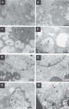
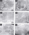
The aim of this study was to evaluate the radioprotector effect of sodium selenite on the ultrastructure of submandibular glands in rats. Fifty-seven male albino Wistar rats were randomized to 4 groups: control, irradiated, sodium selenite and irradiated/sodium selenite. The animals in the sodium selenite and irradiated/sodium selenite groups received intraperitoneal injections of sodium selenite (0.5 mg/kg body weight) 24 h before irradiation. The animals belonging to the irradiated and irradiated/sodium selenite groups were submitted to 15 Gy of gamma radiation in the head and neck region. The submandibular glands were removed at 4, 8, 12, 24, 48 and 72 h after irradiation. The ionizing radiation induced damage to the secretory cells, especially the serous cells, right from the first period. Vacuolization, lysis of cytoplasmic inclusions and nuclear alterations occurred. The sodium selenite group also presented cellular alterations in the study periods, but with less damage compared to that caused by radiation. There was greater similarity between the irradiated/sodium selenite group and the control group than with the other groups treated in all study periods. Despite the alterations observed in the sodium selenite group, sodium selenite presented a radioprotective action on the secretory cells of submandibular glands.
Figures


Similar articles
-
Ultrastructural assessment of the radioprotective effects of sodium selenite on parotid glands in rats.J Oral Sci. 2010 Sep;52(3):369-75. doi: 10.2334/josnusd.52.369. J Oral Sci. 2010. PMID: 20881328
-
[Sodium selenite reduces acute radiogenic damage of the rat parotid glands during fractionated irradiation].HNO. 2004 Dec;52(12):1067-75. doi: 10.1007/s00106-003-0992-x. HNO. 2004. PMID: 15597168 German.
-
Sodium selenite is a potent radioprotector of the salivary glands of the rat: acute effects on the morphology and parenchymal function during fractioned irradiation.Eur Arch Otorhinolaryngol. 2005 Jun;262(6):459-64. doi: 10.1007/s00405-004-0859-0. Epub 2004 Nov 12. Eur Arch Otorhinolaryngol. 2005. PMID: 15942798
-
Effect of sodium selenite and vitamin E on the renal cortex in rats: an ultrastructure study.Tissue Cell. 2014 Jun;46(3):170-7. doi: 10.1016/j.tice.2014.03.002. Epub 2014 Mar 20. Tissue Cell. 2014. PMID: 24799186 Review.
-
Effects of head and neck radiotherapy on major salivary glands--animal studies and human implications.In Vivo. 2003 Jul-Aug;17(4):369-75. In Vivo. 2003. PMID: 12929593 Review.
Cited by
-
Radiation protection following nuclear power accidents: a survey of putative mechanisms involved in the radioprotective actions of taurine during and after radiation exposure.Microb Ecol Health Dis. 2012 Feb 1;23. doi: 10.3402/mehd.v23i0.14787. eCollection 2012. Microb Ecol Health Dis. 2012. PMID: 23990836 Free PMC article.
-
TIGAR overexpression diminishes radiosensitivity of parotid gland fibroblast cells and inhibits IR-induced cell autophagy.Int J Clin Exp Pathol. 2015 May 1;8(5):4823-9. eCollection 2015. Int J Clin Exp Pathol. 2015. PMID: 26191173 Free PMC article.
-
Sodium selenite improves folliculogenesis in radiation-induced ovarian failure: a mechanistic approach.PLoS One. 2012;7(12):e50928. doi: 10.1371/journal.pone.0050928. Epub 2012 Dec 6. PLoS One. 2012. PMID: 23236409 Free PMC article.
-
Radioprotective effects of pyrroloquinoline quinone on parotid glands in C57BL/6J mice.Exp Ther Med. 2016 Dec;12(6):3685-3693. doi: 10.3892/etm.2016.3843. Epub 2016 Oct 26. Exp Ther Med. 2016. Retraction in: Exp Ther Med. 2024 Jul 08;28(3):355. doi: 10.3892/etm.2024.12644. PMID: 28105098 Free PMC article. Retracted.
References
-
- Abok K, Brunk U, Jung B, Ericsson J. Morphologic and histochemical studies on the differing radiosensitivity of ductular and acinar cells of the rat submandibular gland. Virchows Arch B Cell Pathol Incl Mol Pathol. 1984;45:443–460. - PubMed
-
- Ahlner BH, Hagelqvist E, Lind MG. Influence on rabbit submandibular gland injury by stimulation or inhibition of gland function during irradiation-histology and morphometry after 15 gray. Ann Otol Rhinol Laryngol. 1994;103:125–134. - PubMed
-
- Aruoma OI. Characterization of drugs as antioxidant prophylactics. Free Radic Biol Med. 1996;20:675–705. - PubMed
-
- Coppes RP, Vissink A, Konings AWT. Comparison of radiosensitivity of rat parotid and submandibular glands after different radiation schedules. Radiother Oncol. 2002;63:321–328. - PubMed
-
- Delilbasi C, Demiralp S, Turan B. Effects of selenium on the structure of the mandible in experimental diabetics. J Oral Sci. 2002;44:85–90. - PubMed
LinkOut - more resources
Full Text Sources

