

Repair process of surgical defects filled with autogenous bone grafts in tibiae of diabetic rats
- PMID: 19089227
- PMCID: PMC4327596
- DOI: 10.1590/s1678-77572008000500003
Repair process of surgical defects filled with autogenous bone grafts in tibiae of diabetic rats
Abstract
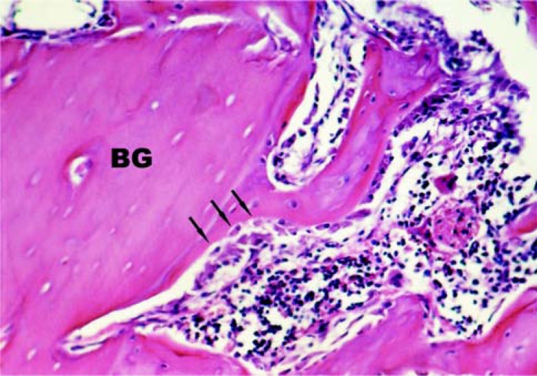
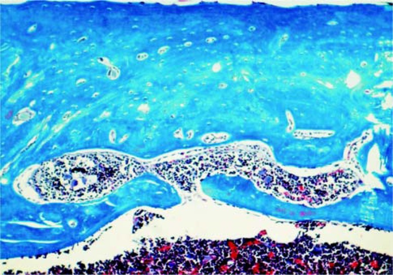
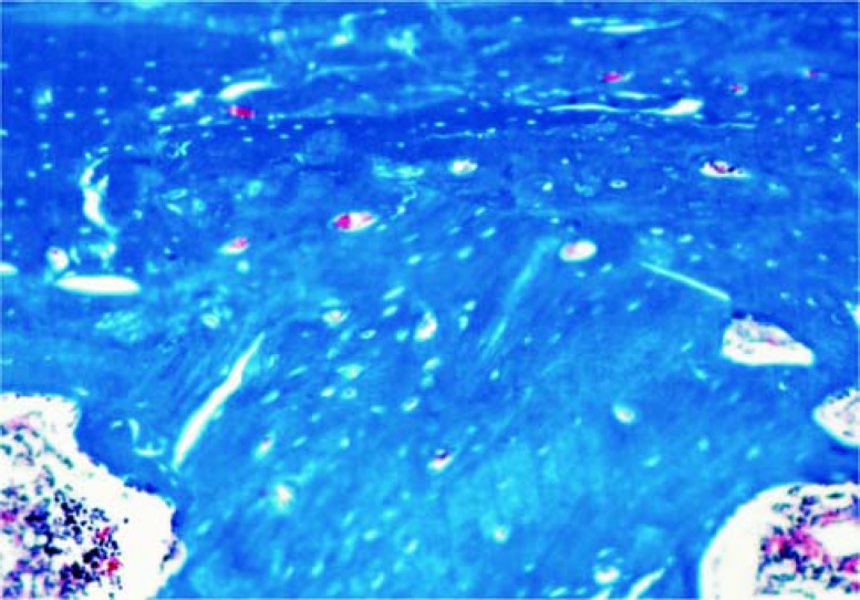
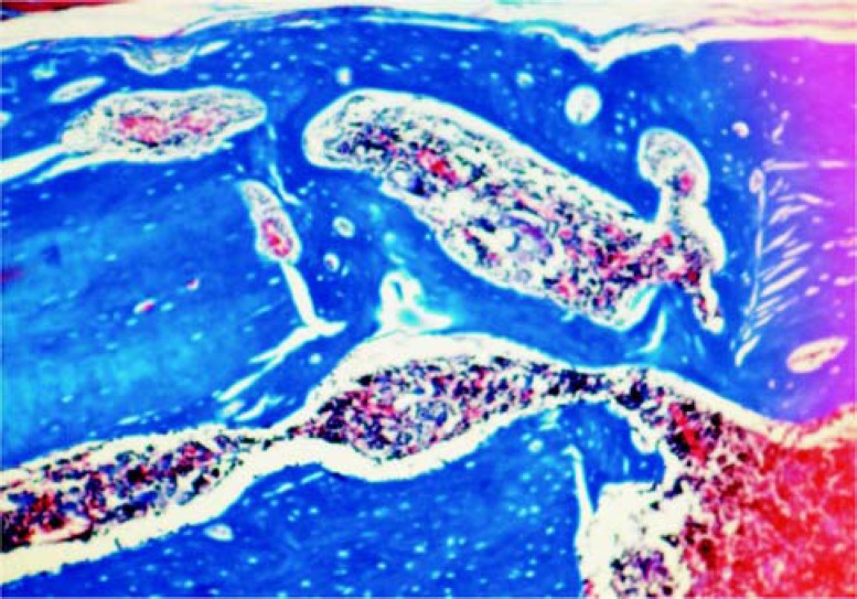
From a biological standpoint, the best material for reconstruction of bone defects is the autogenous bone graft. However, as tissue healing is affected under diabetic conditions, major changes might take place in the revascularization, incorporation, replacement and remodeling phases of the grafted area. The purpose of this study was to assess the bone healing process in surgical wounds prepared in tibiae of diabetic rats and filled with autogenous bone. Forty male rats (Rattus norvegicus albinus, Wistar) were randomly assigned to receive an endovenous injection (penile vein) of either citrate buffer solution (Group 1 - control; n=20) or streptozotocin dissolved in citrate buffer solution (35 mg/kg) to induce diabetes (Group 2 - diabetic; n=20). After determination of glycemia, the animals were anesthetized and the anterolateral regions of the tibiae of both limbs were shaved, antisepsis was performed and longitudinal incisions were made in each limb. The tibiae were exposed and two 2mm-diameter surgical cavities were prepared: one in the right limb, filled with particulate autogenous bone and the other in the left limb, filled with blood clot. The animals were euthanized at 10 and 30 postoperative days. The anatomic pieces were obtained, submitted to laboratory processing and sections were stained by hematoxylin and eosin and Masson's Trichrome for histomorphologic and histometric analyses. In both groups, the wounds filled with autogenous bone graft showed better results than those filled with blood clot. The control group showed higher new bone formation in wounds filled with autogenous bone graft at 30 days than the diabetic group, but without statistical significance. It may be concluded that, in general, the new bone formation occurred with autogenous graft was quantitatively similar between control and diabetic groups and qualitatively better in the control group.
Figures


Similar articles
-
Influence of ovariectomy on healing of autogenous bone block grafts in the mandible: a histomorphometric study in an aged rat model.Int J Oral Maxillofac Implants. 2008 Mar-Apr;23(2):207-14. Int J Oral Maxillofac Implants. 2008. PMID: 18548916
-
Influence of osteopenia in autogenous bone graft healing with or without expanded polytetrafluoroethylene membranes: histologic and histomorphometric study in rats.Int J Oral Maxillofac Implants. 2009 Nov-Dec;24(6):1074-82. Int J Oral Maxillofac Implants. 2009. PMID: 20162112
-
Bone healing in critical-size defects treated with either bone graft, membrane, or a combination of both materials: a histological and histometric study in rat tibiae.Clin Oral Implants Res. 2012 Mar;23(3):384-8. doi: 10.1111/j.1600-0501.2011.02166.x. Epub 2011 Mar 28. Clin Oral Implants Res. 2012. PMID: 21443591
-
Bone healing in critical-size defects treated with platelet-rich plasma: a histologic and histometric study in the calvaria of diabetic rat.Oral Surg Oral Med Oral Pathol Oral Radiol Endod. 2010 Jan;109(1):72-8. doi: 10.1016/j.tripleo.2009.08.003. Epub 2009 Nov 17. Oral Surg Oral Med Oral Pathol Oral Radiol Endod. 2010. PMID: 19926499
-
Bone Repair Process in Defects of Diabetic Rats Filled with Autogenous Bone Graft and Covered with Homogenous Bone Matrix Membrane or Polytetrafluoroethylene Membrane.Int J Oral Maxillofac Implants. 2017 May/June;32(3):e143–e152. doi: 10.11607/jomi.5115. Epub 2017 Mar 23. Int J Oral Maxillofac Implants. 2017. PMID: 28334060
Cited by
-
The subchondral bone in articular cartilage repair: current problems in the surgical management.Knee Surg Sports Traumatol Arthrosc. 2010 Apr;18(4):434-47. doi: 10.1007/s00167-010-1072-x. Epub 2010 Feb 4. Knee Surg Sports Traumatol Arthrosc. 2010. PMID: 20130833 Free PMC article. Review.
-
Bone regeneration in surgically created defects filled with autogenous bone: an epifluorescence microscopy analysis in rats.J Appl Oral Sci. 2010 Jul-Aug;18(4):346-53. doi: 10.1590/s1678-77572010000400005. J Appl Oral Sci. 2010. PMID: 20835568 Free PMC article.
References
-
- Abdulwassie H, Dhanrajani PJ. Diabetes mellitus and dental implants: a clinical study. Implant Dent. 2002;11(1):83–86. - PubMed
-
- Covington DS, Xue H, Pizzini R, Lally KP, Andrassy RJ. Streptozotocin and alloxan are comparable agents in the diabetic model of impaired wound healing. Diabetes Res. 1993;23(2):47–53. - PubMed
-
- Falanga V. Wound healing and its impairment in the diabetic foot. Lancet. 2005;366(9498):1736–1743. - PubMed
Publication types
MeSH terms
Substances
LinkOut - more resources
Full Text Sources
Medical
