Mouth floor enlargements related to the sublingual glands in edentulous or partially edentulous patients: a microscopic study
- PMID: 19089274
- PMCID: PMC4327484
- DOI: 10.1590/s1678-77572006000400010
Mouth floor enlargements related to the sublingual glands in edentulous or partially edentulous patients: a microscopic study
Abstract
Mouth floor enlargements (MFE) are observed in edentulous and partially edentulous patients, impairing denture fitting, and have recently been described in the literature as hyperplasias of the sublingual glands.
Objective: This study aims at describing the microscopic aspects of MFE that contribute to their final diagnosis.
Methods: Twenty-four specimens were surgically removed from the enlarged mouth floor of 19 patients (15 females and 4 males). Patient age ranged from 48 to 74 years, with a mean of 57 years. The main surgical indication was to permit or improve the fitting of dentures. Six patients were completely edentulous and 13 were partially edentulous. The material was processed for microscopic examination and stained with hematoxylin-eosin, Mallory's trichrome and periodic-acid Schiff (PAS).
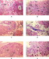
Results and conclusions: The epithelium of the mouth floor was normal in 17 cases, hyperplastic in 4 and atrophic in 3. Six of the 24 sublingual glands removed were microscopically normal, while the other specimens presented acinar atrophy with hyperplasia of duct-like structures. Interstitial fibrosis was observed in 18 cases and was accompanied by adipose tissue infiltration in 15. Decreased lymphoid tissue was observed in 16 samples and oncocytosis was present in 5 cases. We suggest that MFE in edentulous or partially edentulous patients should be considered as an entity for the text books.
Tumefações do soalho bucal (TSB) são observadas em pacientes edêntulos ou parcialmente edêntulos, prejudicando a adaptação de próteses, e têm sido descritas recentemente na literatura como hiperplasias das glândulas sublinguais.
Objetivo:: O objetivo desse estudo é descrever os aspectos microscópicos das TSB a fim de contribuir para o seu diagnóstico final.
Material e Métodos:: Foram removidos cirurgicamente 24 espécimes de 19 pacientes (15 mulheres e 4 homens) que possuíam TSB. A idade variou de 48 a 74 anos, com média de 57 anos. A principal indicação cirúrgica era permitir ou melhorar a adaptação das próteses. Seis pacientes eram edêntulos e 13, parcialmente edêntulos. O material foi processado para exame microscópico e corado com hematoxilina-eosina, tricrômico de Mallory e ácido periódico de Schiff (PAS).
Resultados e Conclusões:: O epitélio do soalho bucal estava normal em 17 casos, hiperplásico em 4 e atrófico em 3. Seis das 24 glândulas sublinguais removidas eram normais microscopicamente, enquanto que as demais apresentaram atrofia acinar com hiperplasia de estruturas ductiformes. Fibrose intersticial foi observada em 18 casos sendo acompanhada por infiltração de tecido adiposo em 15 casos. Uma diminuição no tecido linfóide foi observada em 16 espécimes e oncocitose em 5. Sugerimos que as TSB em pacientes edêntulos e parcialmente edêntulos devem ser classificadas como "entidade" nos livros texto.
Figures


References
-
- Fulop M. Pouting sublinguals: enlarged salivary glands in myxoedema. Lancet. 1989;2:550–551. - PubMed
-
- Tagawa S, Inui M, Mori A, Seki Y, Murata T, Tagawa T. Adenomatoid serous hyperplasia of sublingual gland: a case report. Oral Surg Oral Med Oral Pathol Oral Radiol Endod. 1996;82:437–440. - PubMed
-
- Arafat A, Brannon RB, Ellis GL. Adenomatoid hyperplasia of mucous salivary glands. Oral Surg Oral Med Oral Pathol. 1981;52:51–55. - PubMed
-
- Buchner A, Merrell PW, Carpenter WM, Leider AS. Adenomatoid hyperplasia of minor salivary glands. Oral Surg Oral Med Oral Pathol. 1991;71:583–587. - PubMed
LinkOut - more resources
Full Text Sources

