. 2009:482:215-32.
doi: 10.1007/978-1-59745-060-7_14.

Isolation and culture of epithelial stem cells

Affiliations

Isolation and culture of epithelial stem cells

Jonathan A Nowak et al. Methods Mol Biol. 2009.

Abstract

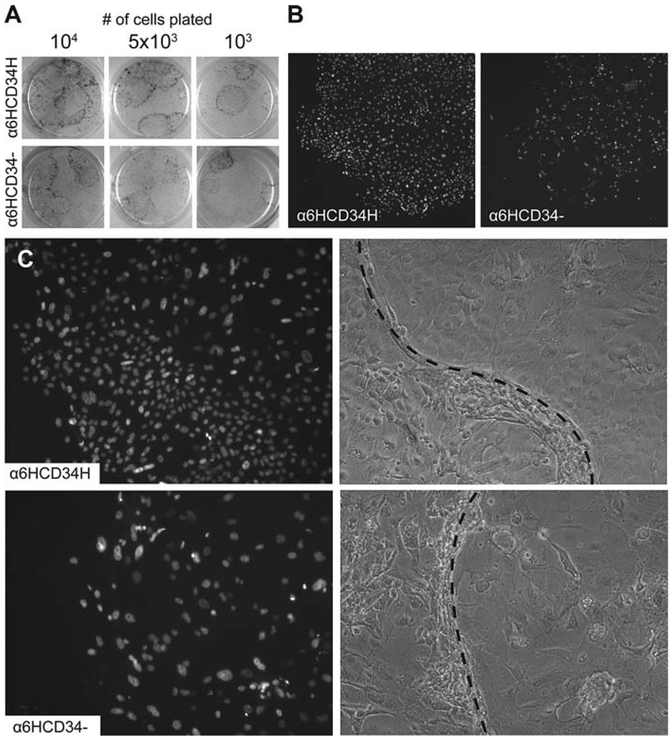
In the skin, epithelial stem cells in the hair follicle contribute not only to the generation of a new hair follicle with each hair cycle, but also to the repair of the epidermis during wound healing. When these stem cells are isolated and expanded in culture, they can give rise to hair follicles, sebaceous glands, and epidermis when combined with dermis and grafted back onto Nude mice. In this chapter, we provide a method for isolating hair follicle epithelial stem cells from the skin of adult mice using immunofluorescent labeling to allow for the specific purification of epithelial stem cells by fluorescence-activated cell sorting (FACS). Notably, this method relies exclusively on cell surface markers, making it suitable for use with any strain of mouse and at various stages of the hair cycle. We also provide a detailed protocol for culturing epithelial stem cells isolated by FACS, allowing for analysis using a wide variety of culture assays. Additionally, we provide notes on using cultured cells for specific applications, such as viral manipulation and grafting. These techniques should be useful for directly evaluating stem cell function in normal mice and in mice with skin defects.


Figures

Fig. 14.1
Fig. 14.1
Identification of hair follicle bulge cells by FACS. Skin epithelial cells from 7-week-old mice were enzymatically isolated to produce a single cell suspension which was stained for α6-integrin and CD34. (A) Protocol used for setting detector voltage and compensation on the flow cytometer. (B) Forward (FSC) versus side scatter (SSC) plot of unstained cells determines the location of the R1 gate, used for excluding debris from all subsequent plots. (C) Unstained cells are visible on the plot of FL2 (PE) and FL4 (APC), but remain within the double negative quadrant. (D,F) Singly stained α6-PE and CD34-APC control cells distribute along the expected axes. (E) Propidium iodide (PI) staining is used to identify viable cells, shown here in region R2. (G) Double staining for CD34-APC and α6-PE, along with gating on regions R1 and R2, demonstrates two distinct bulge cell populations—the suprabasal α6LowCD34High and basal α6HighCD34High population.
Fig. 14.2
Fig. 14.2
Expression levels of α6-integrin distinguish two populations of CD34High bulge cells. (A) CD34 is specifically enriched in the bulge region of the hair follicle beginning at the first telogen stage of the hair cycle, approximately day 20. A single layer of uniformly α6High basal bulge cells (dark grey) surround the lower portion of the club hair, below the sebaceous gland (SG). Basal cells of the interfollicular epidermis (IFE) and upper hair follicle are present as a α6HighCD34 population. (B) During the next anagen stage of the hair cycle, the growth of a second hair shaft creates a new population of α6LowCD34High suprabasal bulge cells (light grey). During this stage, cells from the lower, cycling portion of the outer root sheath (ORS) are also visible as a population of α6LowCD34Low/− cells. (C) During the second telogen stage of the hair cycle, both the α6HighCD34High and α6LowCD34High populations are distinct, and the cycling portion of the outer root sheath is absent.
Fig. 14.3
Fig. 14.3
Bulge cells display distinct growth properties when placed in culture. The α6HighCD34High bulge cells and α6HighCD34 epidermal cells were isolated by FACS from mice expressing a GFP-tagged histone H2B under the control of a keratin 14 promoter. (A) Rhodamine B staining of cultured cells shows that although both cell populations have similar colony forming efficiencies, colonies derived from bulge cells tend to be larger and more regularly shaped. (B) GFP fluorescence demonstrates that α6HighCD34High cells give rise to densely packed colonies with regular borders, while α6HighCD34 cells generate sparser colonies with a more irregular appearance. (C) GFP fluorescence (left) and phase contrast images (right) show that colonies derived from α6HighCD34High cells consist largely of small, undifferentiated cells with a uniform morphology, while α6HighCD34 colonies contain a mix of larger cells with more variable morphology. Note that colonies begin growing underneath the feeder cell layer, and feeder cells are gradually pushed aside as the colony expands in size. Dotted lines indicate the approximate boundary between feeder cells (left) and epidermal cells (right).

References

    1. Cotsarelis G, Sun TT, Lavker RM. Label-retaining cells reside in the bulge area of pilosebaceous unit: implications for follicular stem cells, hair cycle, and skin carcinogenesis. Cell. 1990;61(7):1329–1337. - PubMed
    1. Morris RJ, Potten CS. Highly persistent label-retaining cells in the hair follicles of mice and their fate following induction of anagen. J Invest Dermatol. 1999;112(4):470–475. - PubMed
    1. Taylor G, et al. Involvement of follicular stem cells in forming not only the follicle but also the epidermis. Cell. 2000;102(4):451–461. - PubMed
    1. Tumbar T, et al. Defining the epithelial stem cell niche in skin. Science. 2004;303(5656):359–363. - PMC - PubMed
    1. Morris RJ, et al. Capturing and profiling adult hair follicle stem cells. Nat Biotechnol. 2004;22(4):411–417. - PubMed