

Isolation and characterization of hepatic stem cells, or "oval cells," from rat livers
- PMID: 19089369
- PMCID: PMC3130598
- DOI: 10.1007/978-1-59745-060-7_24
Isolation and characterization of hepatic stem cells, or "oval cells," from rat livers
Abstract
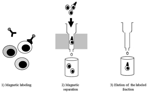
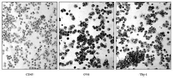
The pace of research on the potential therapeutic uses of liver stem cells or "oval cells" has accelerated significantly in recent years. Concurrent advancements in techniques for the isolation and characterization of these cells have helped fuel this research. Several models now exist for the induction of oval cell proliferation in rodents. Protocols for the isolation and culture of these cells have evolved to the point that they may be set up in any laboratory equipped for cell culture. The advent of magnetic cell sorting has eliminated reliance on expensive flow cytometric sorting equipment to generate highly enriched populations of oval cells. Our laboratory has had much success in using the oval cell surface marker Thy-1 in combination with magnetic sorting to produce material suitable for testing the influence of a myriad of chemical signaling molecules on the oval cell phenotype. This chapter will describe our basic strategy for oval cell induction and isolation. Additionally, two in vitro procedures are described which the reader may find useful in the early stages of developing an oval cell research project.
Figures
Similar articles
-
Characterization and enrichment of hepatic progenitor cells in adult rat liver.World J Gastroenterol. 2004 May 15;10(10):1480-6. doi: 10.3748/wjg.v10.i10.1480. World J Gastroenterol. 2004. PMID: 15133858 Free PMC article.
-
Activation, isolation, identification and in vitro proliferation of oval cells from adult rat livers.Cell Prolif. 2004 Apr;37(2):177-87. doi: 10.1111/j.1365-2184.2004.00293.x. Cell Prolif. 2004. PMID: 15030551 Free PMC article.
-
Isolation of hepatic progenitor cells from the galactosamine-treated rat liver.Methods Mol Biol. 2012;826:49-58. doi: 10.1007/978-1-61779-468-1_5. Methods Mol Biol. 2012. PMID: 22167639
-
Hepatic progenitor populations in embryonic, neonatal, and adult liver.Proc Soc Exp Biol Med. 1993 Dec;204(3):261-9. doi: 10.3181/00379727-204-43662. Proc Soc Exp Biol Med. 1993. PMID: 8234369 Review.
-
[Identification and enrichment of hepatic stem cells by flow cytometric cell sorting].Tanpakushitsu Kakusan Koso. 2000 Sep;45(13 Suppl):2250-6. Tanpakushitsu Kakusan Koso. 2000. PMID: 11021231 Review. Japanese. No abstract available.
Cited by
-
Unbalanced distribution of materials: the art of giving rise to hepatocytes from liver stem/progenitor cells.J Cell Mol Med. 2014 Jan;18(1):1-14. doi: 10.1111/jcmm.12183. Epub 2013 Nov 28. J Cell Mol Med. 2014. PMID: 24286303 Free PMC article. Review.
-
Advances in Hepatic Tissue Bioengineering with Decellularized Liver Bioscaffold.Stem Cells Int. 2019 May 6;2019:2693189. doi: 10.1155/2019/2693189. eCollection 2019. Stem Cells Int. 2019. PMID: 31198426 Free PMC article. Review.
-
Directed Differentiation of Adult Liver Derived Mesenchymal Like Stem Cells into Functional Hepatocytes.Sci Rep. 2018 Feb 12;8(1):2818. doi: 10.1038/s41598-018-20304-5. Sci Rep. 2018. PMID: 29434311 Free PMC article.
-
A label-retaining but unipotent cell population resides in biliary compartment of mammalian liver.Sci Rep. 2017 Jan 13;7:40322. doi: 10.1038/srep40322. Sci Rep. 2017. PMID: 28084309 Free PMC article.
-
Stem cell origins and animal models of hepatocellular carcinoma.Dig Dis Sci. 2010 May;55(5):1241-50. doi: 10.1007/s10620-009-0861-x. Epub 2009 Jun 10. Dig Dis Sci. 2010. PMID: 19513833 Review.
References
-
- Newsome PN, Hussain MA, Theise ND. Hepatic oval cells: helping redefine a paradigm in stem cell biology. Curr Top Dev Biol. 2004;61:1–28. - PubMed
-
- Oh SH, Hatch HM, Petersen BE. Hepatic oval ‘stem’ cell in liver regeneration. Semin Cell Dev Biol. 2002;13:405–409. - PubMed
-
- Petersen BE. Hepatic “stem” cells: coming full circle. Blood Cells Mol Dis. 2001;27:590–600. - PubMed
-
- Piscaglia AC, Di Campli C, Gasbarrini G, Gasbarrini A. Stem cells: new tools in gastroenterology and hepatology. Dig Liver Dis. 2003;35:507–514. - PubMed
-
- Lowes KN, Croager EJ, Olynyk JK, Abraham LJ, Yeoh GC. Oval cell-mediated liver regeneration: Role of cytokines and growth factors. J Gastroenterol Hepatol. 2003;18:4–12. - PubMed
MeSH terms
Substances
Grants and funding
LinkOut - more resources
Full Text Sources
Other Literature Sources
Medical
Miscellaneous
