Review
. 2009 Feb;87(2):125-38.
doi: 10.1007/s00109-008-0426-z. Epub 2008 Dec 17.

Toll-like receptors, wound healing, and carcinogenesis

Affiliations
Review

Toll-like receptors, wound healing, and carcinogenesis

Johannes Kluwe et al. J Mol Med (Berl). 2009 Feb.

Abstract

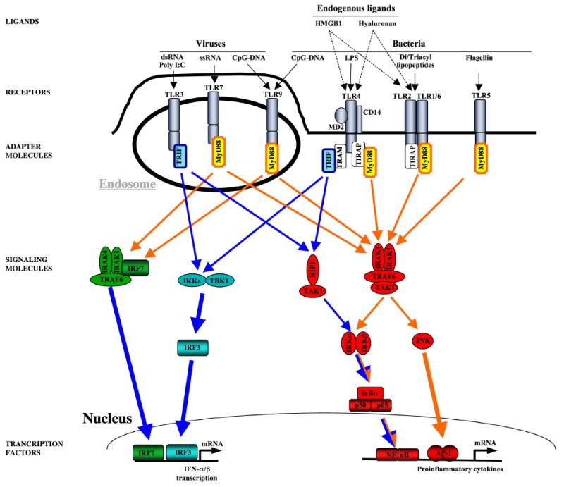
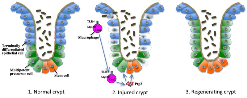
Following acute injury, the concerted action of resident and nonresident cell populations evokes wound healing responses that entail a temporary increase in inflammation, extracellular matrix production, and proliferation to ultimately restore normal organ architecture. However, chronic injury evokes a perpetuating wound healing response promoting the development of fibrosis, organ failure, and cancer. Recent evidence points toward toll-like receptors (TLRs) as important regulators of inflammatory signals in wound healing. Here, we will review the activation of TLRs by different endogenous and bacterial TLR ligands during wound healing, and the contribution of TLR-induced signals to injury, fibrogenesis, regeneration, and carcinogenesis. We will discuss the hypothesis that TLRs act as sensors of danger signals in injured tissue to switch the wound healing response toward fibrogenesis and regeneration as a protective response to imminent danger at the cost of an increased long-term risk of developing scars and cancer.


Figures

Figure 1
Figure 1. TLR signaling
Viral PAMPS activate TLR3, TLR7, and TLR9, whereas bacterial PAMPs activate TLR2/1, TLR2/6, TLR4, and TLR9. Several endogenous mediators including hyaluronan and HMGB1 have been suggested to activate TLR2 and TLR4. TLRs mediate their signaling through to adapater molecules, MyD88 and TRIF to induce up-regulation proinflammatory and antiviral genes. MyD88-induced signals (marked in orange) predominantly activate NF-κB, IRF-7 and JNK, Trif-dependent signals (marked in blue) predominantly activate NF-κB and IRF-3.
Figure 2
Figure 2. TLR-mediated epithelial regeneration after DSS-mediated colonic injury
In the colonic crypt, stem cells generate multipotent precursor cells which then differentiate into different types of terminally differentiated epithelial cell including enteroabsorptive, paneth and goblet cells to chronically replace the epithelial layer of crypts. Following DSS-induced injury, terminally differentiated epithelial cells as well as multipotent precursor cells undergo cell death. TLR4- and MyD88-dependent signals are required to induce Cox2-mediated generation of prostaglandin2 (ptg2) and to stimulate epithelial cell proliferation. The MyD88-Cox2 signal that promotes regeneration is largely provided by macrophages which migrate towards the site of injury to stimulate proliferation in Cox2-dependent manner. (Figure adapted from Rakoff-Nahoum et al. [82].
Figure 3
Figure 3. Mechanisms by which TLR4 promotes hepatic stellate cell activation and liver fibrosis
Changes in the intestinal motility, mucosa and microbiota promote release of LPS from the intestinal microbiota. These relatively low amounts of LPS do not induce any appreciable liver injury but target hepatic stellate cells to downregulate TGFβ pseudoreceptor Bambi and upregulate chemokines. These two signals work hand in hand to promote hepatic stellate cell activation: 1. Downregulation of Bambi through a TLR4-MyD88-NF-κB signaling cascade sensitizes hepatic stellate cells towards the effect of TGFβ. 2. Chemokines induce Kupffer cells, a main source of TGFβ in the injured liver, to migrate towards hepatic stellate cells. Thus, the LPS-TLR4-MyD88 signaling cascade mediates the cross-talk between HSCs and Kupffer cells resulting in unrestricted TGFβ-mediated activation of hepatic stellate cells, increased deposition of extracellular matrix and promotion of liver fibrosis.

References

    1. Bataller R, Brenner DA. Liver fibrosis. J Clin Invest. 2005;115:209–218. doi: 10.1172/JCI24282. - DOI - PMC - PubMed
    1. Liu Y. Renal fibrosis: new insights into the pathogenesis and therapeutics. Kidney Int. 2006;69:213–217. doi: 10.1038/sj.ki.5000054. DOI 5000054 [pii] - DOI - PubMed
    1. Martinez FJ, Safrin S, Weycker D, Starko KM, Bradford WZ, King TE, Jr, Flaherty KR, Schwartz DA, Noble PW, Raghu G, Brown KK. The clinical course of patients with idiopathic pulmonary fibrosis. Ann Intern Med. 2005;142:963–967. DOI 142/12_Part_1/963 [pii] - PubMed
    1. Swynghedauw B. Molecular mechanisms of myocardial remodeling. Physiol Rev. 1999;79:215–262. - PubMed
    1. Dvorak HF. Tumors: wounds that do not heal. Similarities between tumor stroma generation and wound healing. N Engl J Med. 1986;315:1650–1659. - PubMed

Publication types

Substances

LinkOut - more resources