. 2009 Apr 1;124(7):1545-51.
doi: 10.1002/ijc.24118.

Downregulation of vascular endothelial growth factor and induction of tumor dormancy by 15-lipoxygenase-2 in prostate cancer

Affiliations

Downregulation of vascular endothelial growth factor and induction of tumor dormancy by 15-lipoxygenase-2 in prostate cancer

Yong Tang et al. Int J Cancer. .

Abstract

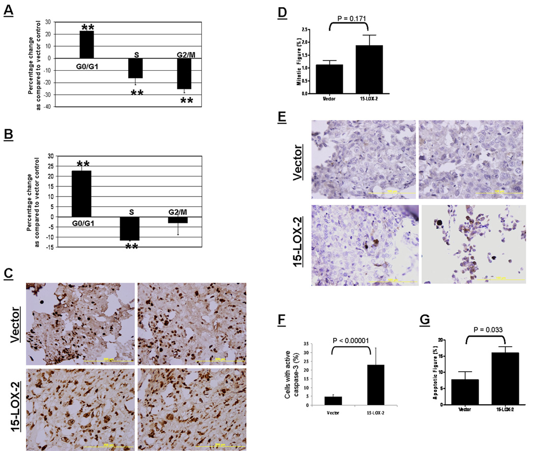
The enzyme 15-lipoxygenase-2 (15-LOX-2) utilizes arachidonic acid, a polyunsaturated fatty acid, to synthesize 15(S)-hydroxyeicosatetraenoic acid. Abundantly expressed in normal prostate epithelium but frequently suppressed in the cancerous tissues, 15-LOX-2 has been suggested as a functional suppressor of prostate cancer, but the mechanism(s) involved remains unknown. To study the functional role of 15-LOX-2 in prostate cancer, we expressed 15-LOX-2 as a fusion protein with GFP in DU145 and PC-3 cells and found that 15-LOX-2 increased cell cycle arrest at G0/G1 phase. When injected into athymic nu/nu mice, prostate cancer cells with 15-LOX-2 expression could still form palpable tumors without significant changes in tumorigenicity. But, the tumors with 15-LOX-2 expression grew significantly slower than those derived from vector controls and were kept dormant for a long period of time. Histological evaluation revealed an increase in cell death in tumors derived from prostate cancer cells with 15-LOX-2 expression, while in vitro cell culture conditions, no such increase in apoptosis was observed. Further studies found that the expression of vascular endothelial growth factor A (VEGF-A) was significantly reduced in prostate cancer cells with 15-LOX-2 expression restored. Our studies suggest that 15-LOX-2 suppresses VEGF gene expression and sustains tumor dormancy in prostate cancer. Loss of 15-LOX-2 functionalities, therefore, represents a key step for prostate cancer cells to exit from dormancy and embark on malignant progression in vivo.


Figures

Figure 1
Figure 1. Expression and activity of 15-LOX-2 in prostate cancer cells
A. Expression of 15-LOX-2 as a fusion protein with GFP in PC-3 cells. Upper panel, the blot probed for 15-LOX-2 expression. Bottom panel, the same blot stripped and reprobed for GFP. Note the expression of 15-LOX-2/GFP fusion protein with estimated molecular weight around 112 kD. B. Analysis of 15(S)-HETE by HPLC-tandem MS. Sample extracts from ~6 million PC-3 cells were analyzed. Cells transfected with 15-LOX-2 (top) had ~100 fold increase in 15(S)-HETE compared to the control vector (bottom). 12(S)-HETE d8 is the internal standard used for quantitation and is kept at a constant level.
Figure 2
Figure 2. Induction of prostate tumor dormancy in vivo by 15-LOX-2
A. Six-week growth kinetics of the tumors derived from DU145 15-LOX-2 transfectants and vector control (GFP). Data point, mean volume of six tumors for 15-LOX-2 transfectants and five tumors for the vector controls; bars, STD. B. Mice with tumors and gross morphology of tumors from PC-3 15-LOX-2 transfectants or from vector controls. Top panel, mice with tumors in situ; bottom panel, gross morphology of tumors resected.
Figure 3
Figure 3. Mechanism of tumor dormancy by 15-LOX-2
A. Increased G0/G1 arrest in DU145 cells by 15-LOX-2 in serum free media. Vector control and 15-LOX-2 transfectants were plated and cultured in serum free media for 24 h and then the cells were trypsinized, fixed in 70% ethanol, and processed for flow cytometry analysis of cell cycle and processed for flow cytometry analysis of cell cycle. The percentage changes in each phase were calculated by comparing to the vector control. Values are the average of percentage changes from the three samples ± STD. **, P < 0.01 between 15-LOX-2 transfectants and the vector control. B. Increased G0/G1 arrest in DU145 cells by 15-LOX-2 expression in serum containing media. Vector control and 15-LOX-2 transfectants were cultured in RPMI-10% FBS for 24 h and then the cells were harvested and processed for cell cycle analysis as described above. Values are the average of the percentage changes from three repeats ± STD. **, P < 0.01 between 15-LOX-2 transfectants and the vector control. C. Ki-67 staining of proliferating cells. The proliferating cells in tumor sections were revealed by Ki-67 staining using standard IHC protocol (Brown staining). Note the lack of reduction in cell proliferation in tumors derived from 15-LOX-2 transfected PC-3 cells (Bottom panel) when compared to those from the vector controls (Upper panel). D. Mitotic index. Mitotic figures were counted in 400 X fields and expressed as the percentage of the total cells. Columns, mean mitotic index in tumors; bars, STD. Note the slight but not statistically significant increase in mitotic figures in tumors derived from 15-LOX-2 transfectants (n = 5) as compared to those of vector controls (n = 4) (P > 0.05 by Student’s t test). E. Increased caspase-3 activation in tumors from 15-LOX-2 transfected cells. The active form of caspase-3 in tumor sections was evaluated by IHC staining using an antibody against cleaved caspase-3 using standard ABC protocol with hematoxylin counter staining. Brown color indicates positive staining. F. Percentage of cells with caspase-3 activation. Tumor sections stained with cleaved caspase-3 were observed and digital pictures were taken at 200X magnification. Positively stained cells were counted and expressed as the percentage of the total cells in the fields. Column, average percentage of cells positive for cleaved caspase-3; Bars, STD. n = 10 for the vector group. For 15-LOX-2 group, n = 11. G. Apoptotic index. Apoptotic figures were counted in 400 X fields and the index expressed as the percentage of the total cells in the same field. Columns, mean apoptotic index in tumors; bars, STD. Note the significant increase in apoptotic figures in tumors derived from 15-LOX-2 transfectants (n =5) as compared to those of vector controls (n = 4) (P < 0.05 by Student’s t test).
Figure 4
Figure 4. Down-regulation of VEGF expression in prostate cancer cells by 15-LOX-2
A. Reduced VEGF production in DU145 and PC-3 cells as result of 15-LOX-2 expression. The levels of VEGF in the conditioned media from 15-LOX-2 transfected prostate cancer cells (PC-3 and DU145) and their respective vector control cells were determined using ELISA. The values were normalized with respective vector controls, which were set as 100%. Columns, mean values of three samples; bars, STD. *, p < 0.05. B. Reduced immunoreactivities of VEGF in tumor sections derived from PC-3 15-LOX-2 transfectants. The negative control (top panel) is the tumor sections incubated with reagents except VEGF antibody as quality control for IHC. The VEGF immunoreactivities were indicated by the dark brown staining. Shown here are two representative tumor sections from four different tumors. C. Reduced VEGF promoter activities in DU145 cells stably transfected with 15-LOX-2, but not by other isoforms of LOX, 15-LOX-1 or 5-LOX. D. Reduction of VEGF promoter (p− 1176/+45) activities in PC-3 cells transfected with 15-LOX-2. Cells were transfected with a VEGF promoter luciferase construct (− 1176/+54) and a LacZ β-gal expression construct as described in Materials and Methods. Values are the average of normalized luciferase activities from three dishes ± STD. P values were calculated by comparing to the vector control (GFP) (Student’s t test).

References

    1. Jemal A, Siegel R, Ward E, Murray T, Xu J, Smigal C, Thun MJ. Cancer statistics, 2006. CA Cancer J Clin. 2006;56:106–130. - PubMed
    1. Folkman J, Kalluri R. Cancer without disease. Nature. 2004;427:787. - PubMed
    1. Sakr WA, Ward C, Grignon DJ, Haas GP. Epidemiology and molecular biology of early prostatic neoplasia. Mol Urol. 2000;4:109–113. discussion 15. - PubMed
    1. Brash AR, Boeglin WE, Chang MS. Discovery of a second 15S-lipoxygenase in humans. Proc Natl Acad Sci U S A. 1997;94:6148–6152. - PMC - PubMed
    1. Shappell SB, Boeglin WE, Olson SJ, Kasper S, Brash AR. 15-lipoxygenase-2 (15-LOX-2) is expressed in benign prostatic epithelium and reduced in prostate adenocarcinoma. Am J Pathol. 1999;155:235–245. - PMC - PubMed

Publication types

MeSH terms

Substances