. 2009 Jun;75(4):931-53.
doi: 10.1002/prot.22304.

Mechanism of formation of the C-terminal beta-hairpin of the B3 domain of the immunoglobulin binding protein G from Streptococcus. I. Importance of hydrophobic interactions in stabilization of beta-hairpin structure

Affiliations

Mechanism of formation of the C-terminal beta-hairpin of the B3 domain of the immunoglobulin binding protein G from Streptococcus. I. Importance of hydrophobic interactions in stabilization of beta-hairpin structure

Agnieszka Skwierawska et al. Proteins. 2009 Jun.

Abstract

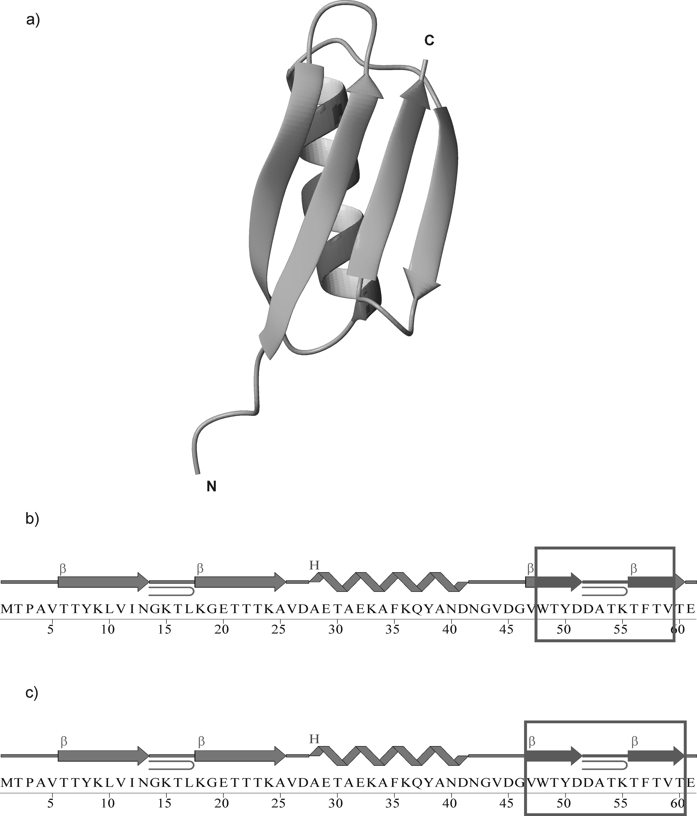
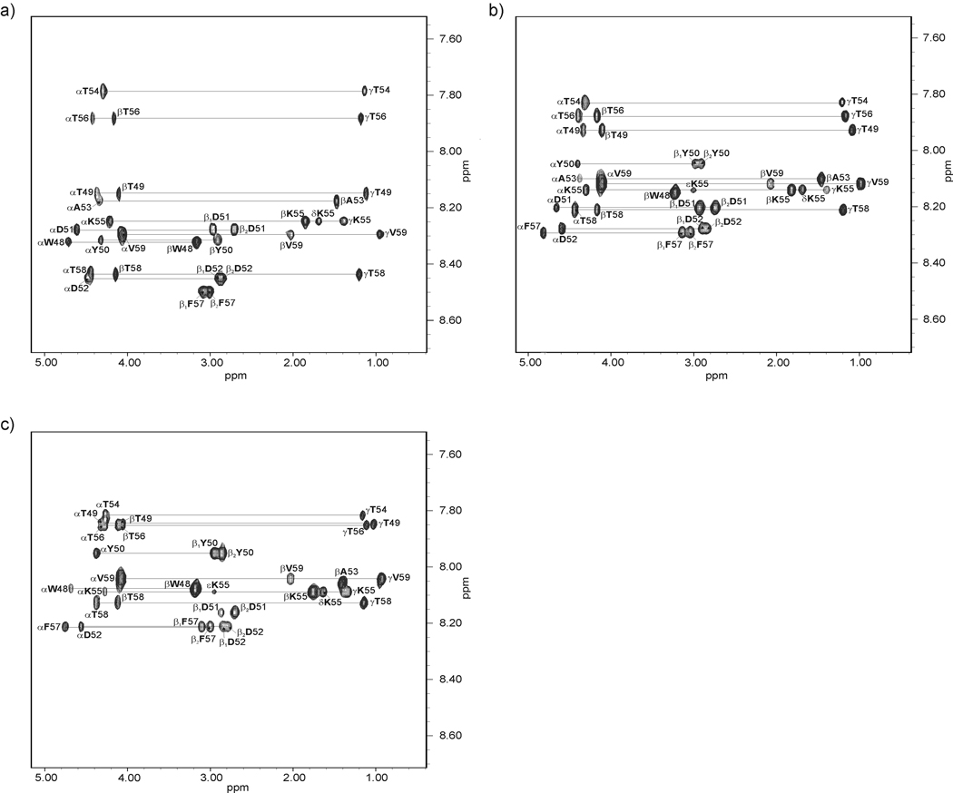
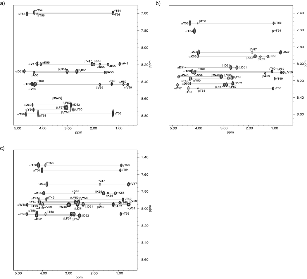
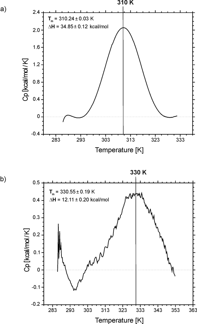
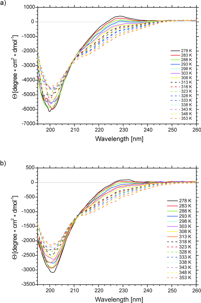
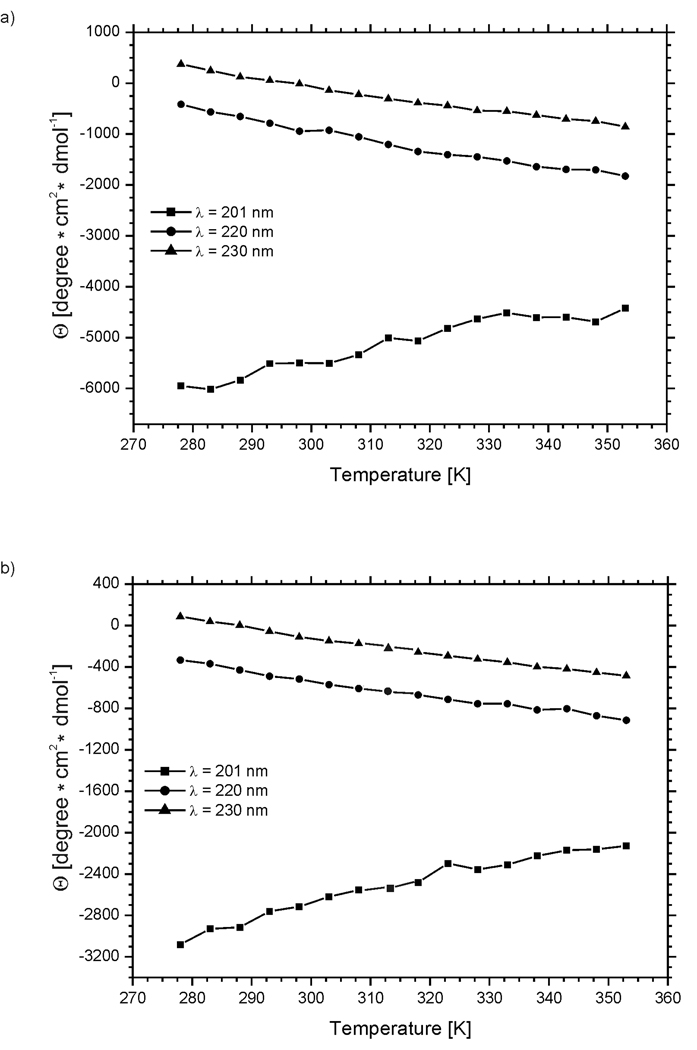
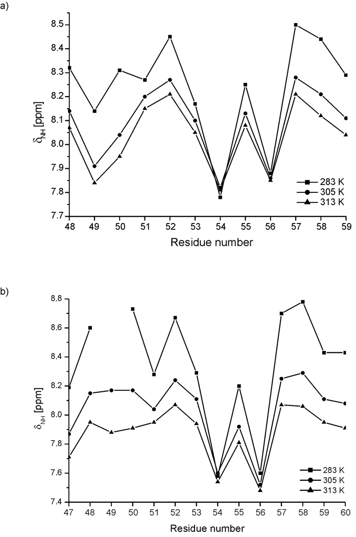
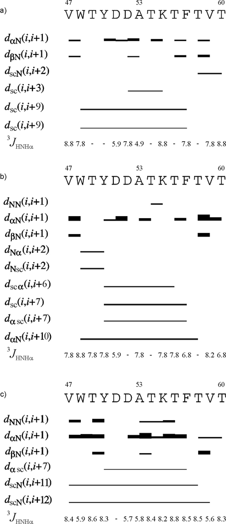
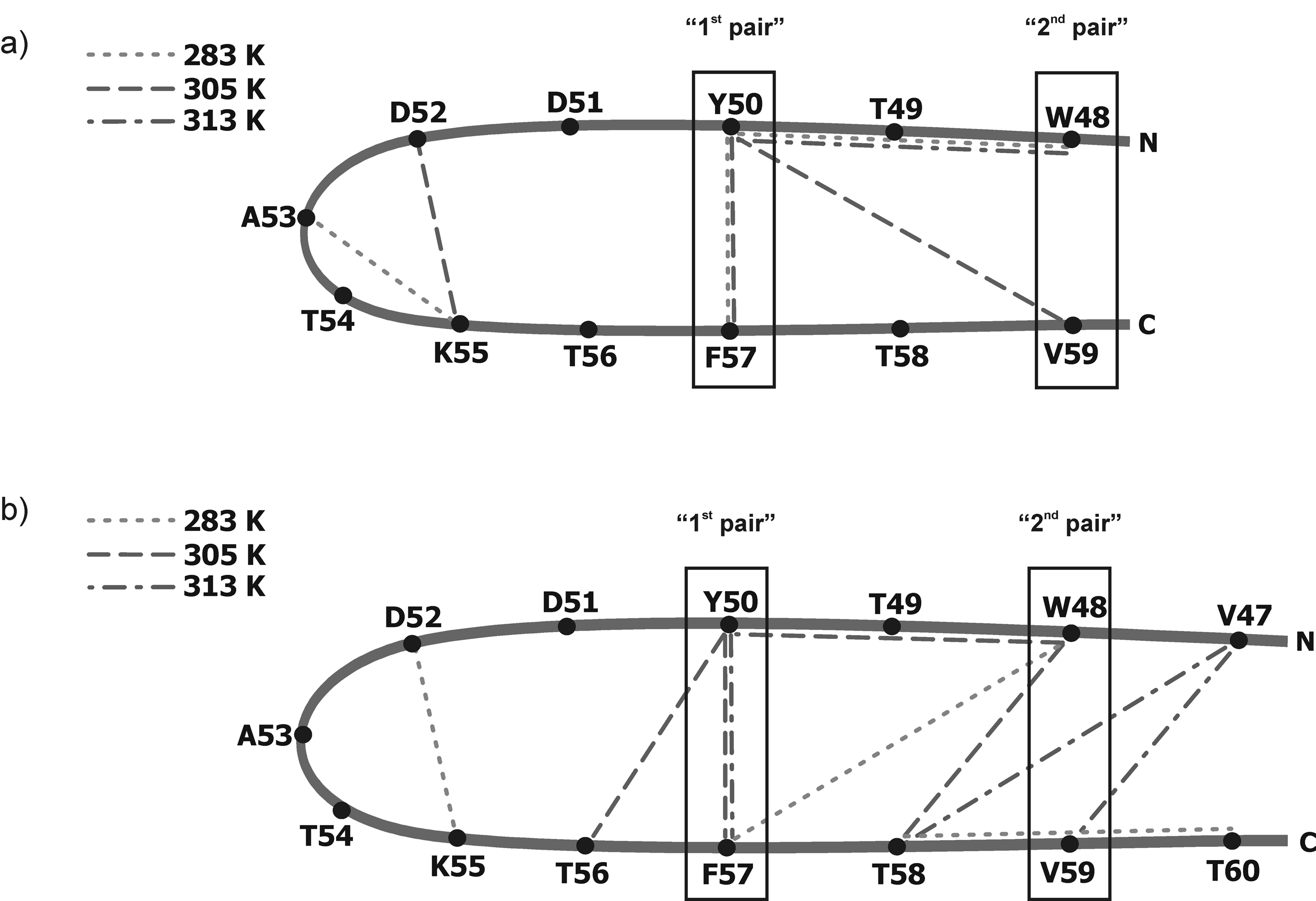
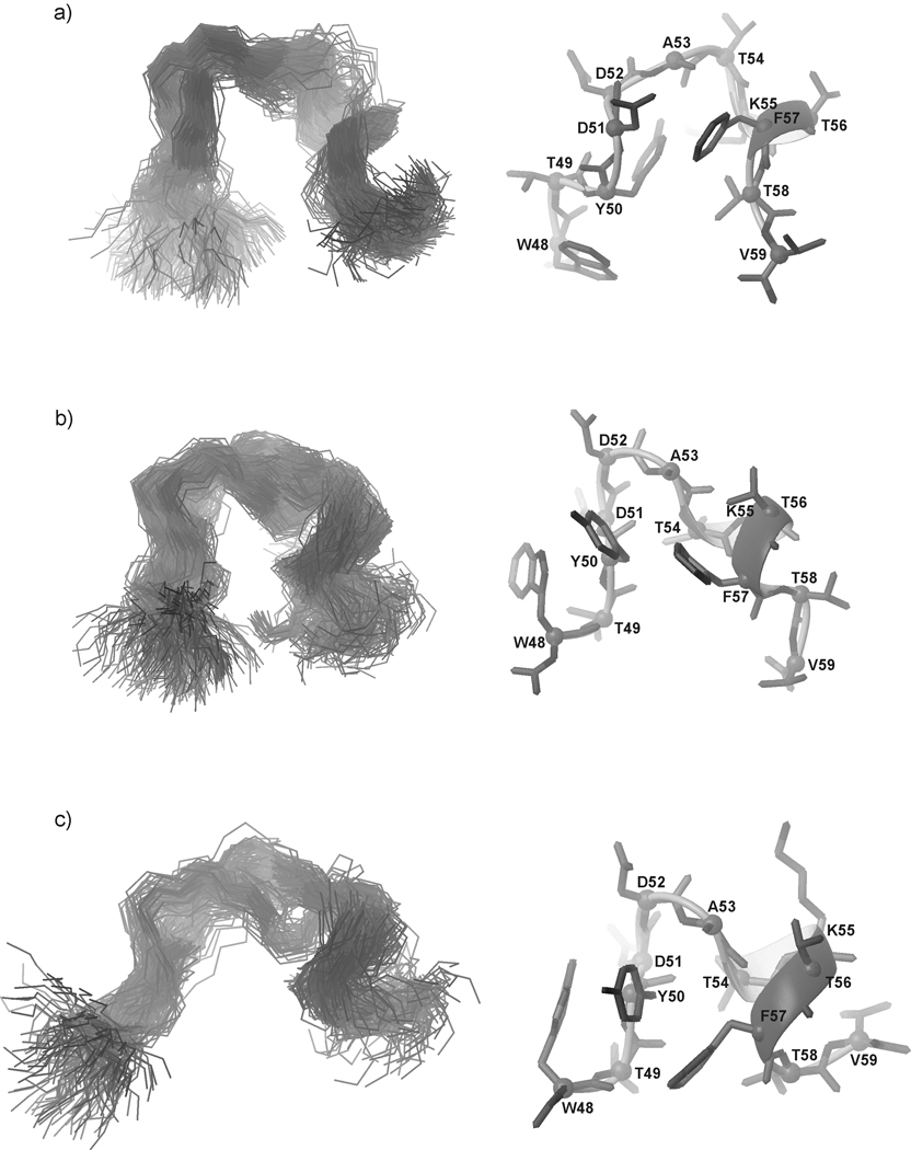
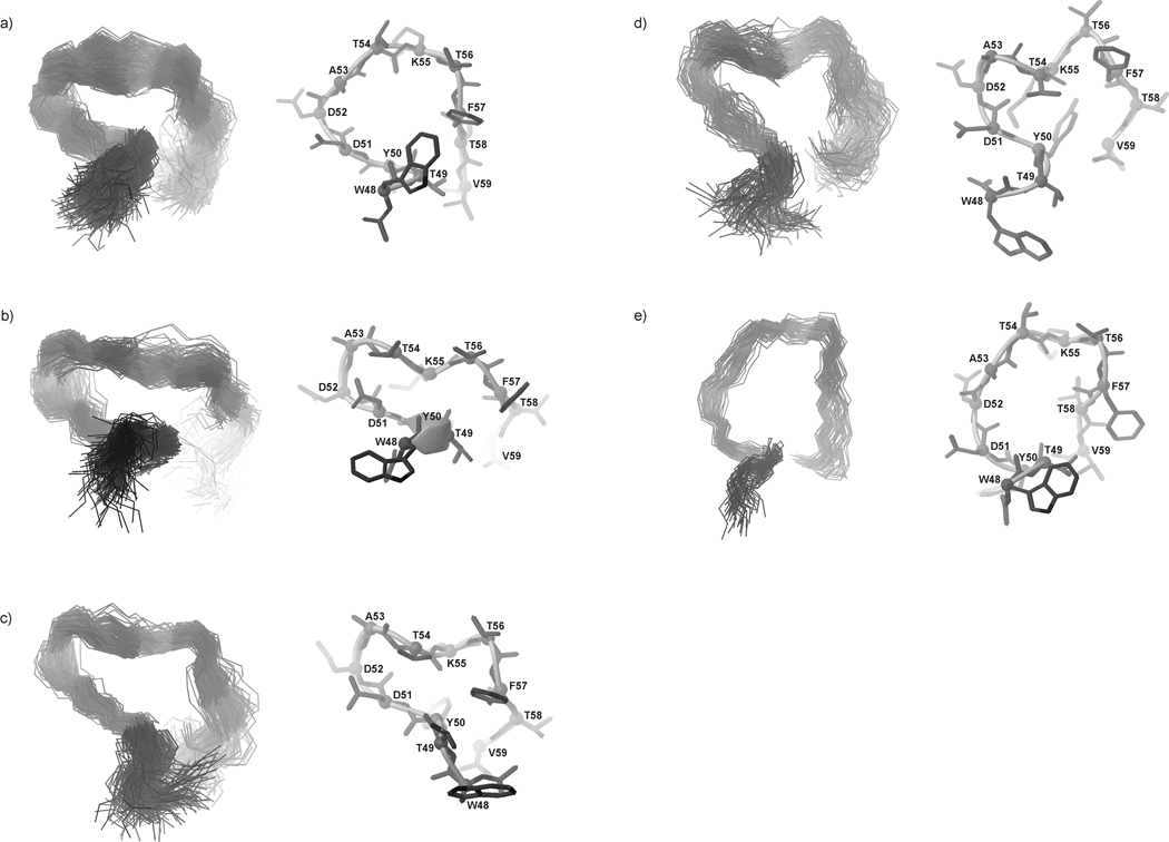
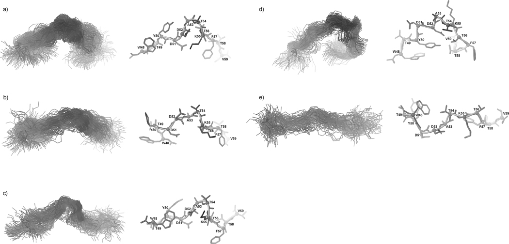
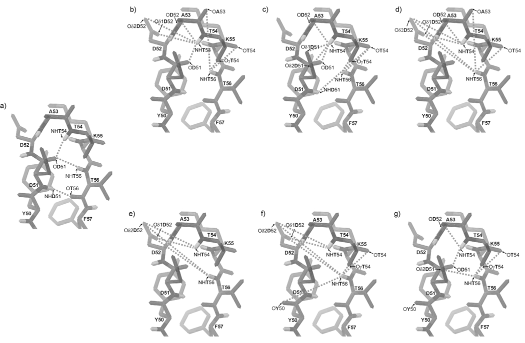
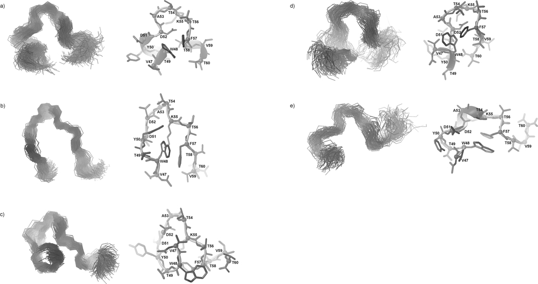
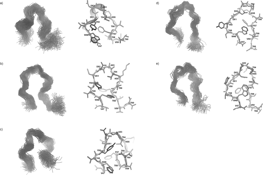
We previously studied a 16-amino acid-residue fragment of the C-terminal beta-hairpin of the B3 domain (residues 46-61), [IG(46-61)] of the immunoglobulin binding protein G from Streptoccocus, and found that hydrophobic interactions and the turn region play an important role in stabilizing the structure. Based on these results, we carried out systematic structural studies of peptides derived from the sequence of IG (46-61) by systematically shortening the peptide by one residue at a time from both the C- and the N-terminus. To determine the structure and stability of two resulting 12- and 14-amino acid-residue peptides, IG(48-59) and IG(47-60), respectively, we carried out circular dichroism, NMR, and calorimetric studies of these peptides in pure water. Our results show that IG(48-59) possesses organized three-dimensional structure stabilized by hydrophobic interactions (Tyr50-Phe57 and Trp48-Val59) at T = 283 and 305 K. At T = 313 K, the structure breaks down because of increased chain entropy, but the turn region is preserved in the same position observed for the structure of the whole protein. The breakdown of structure occurs near the melting temperature of this peptide (T(m) = 310 K) measured by differential scanning calorimetry (DSC). The melting temperature of IG(47-60) determined by DSC is T(m) = 330 K and its structure is similar to that of the native beta-hairpin at all (lower) temperatures examined (283-313 K). Both of these truncated sequences are conserved in all known amino acid sequences of the B domains of the immunoglobulin binding protein G from bacteria. Thus, this study contributes to an understanding of the mechanism of folding of this whole family of proteins, and provides information about the mechanism of formation and stabilization of a beta-hairpin structural element.


Figures

Fig. 1
Fig. 1
X-ray structure of the B3 domain of protein G (PDB nomenclature: 1IGD) (a); Amino acid sequence of 1IGD, where the boxed fragments, IG(48–59) (b), and IG(47–60) (c), were synthesized and examined. In (b) and (c), β and H denote strand and helix, respectively.
Fig. 2
Fig. 2
Amino acid spin systems in the TOCSY spectra of IG(48–59) in H2O at pH = 5.13 (tm=80 ms; the diagnostic region) at (a) 283 K, (b) 305 K and (c) 313 K.
Fig. 3
Fig. 3
Amino acid spin systems in the TOCSY spectra of IG(47–60) in H2O at pH = 5.45 (tm=80 ms; the diagnostic region) at (a) 283 K, (b) 305 K and (c) 313 K.
Fig. 4
Fig. 4
Heat capacity curves for (a) IG(48–59) and (b) IG(47–60) recorded in water at pH = 5.13 and 5.45, respectively.
Fig. 5
Fig. 5
CD spectra of (a) IG(48–59) and (b) IG(47–60) in water at 16 different temperatures.
Fig. 6
Fig. 6
Variation of the molar ellipticity of (a) IG(48–59) and (b) IG(47–60), at 201 nm (squares), 220 nm (circles), and 230 nm (triangles), respectively, with temperature.
Fig. 7
Fig. 7
Chemical shifts of the amide protons of consecutive amino acid residues of (a) IG(48–59) and (b) IG(47–60) at pH = 5.13 and 5.45, respectively, at three different temperatures (283, 305 and 313 K).
Fig. 8
Fig. 8
ROE effects corresponding to the interproton contacts and the 3JNHHα coupling constants of IG(48–59) measured in H2O at (a) 283 K, (b) 305 K and (c) 313 K. The thickness of the bars reflects the strength of the ROE correlation as strong, medium or weak.
Fig. 9
Fig. 9
ROE effects corresponding to the interproton contacts and the 3JNHHα coupling constants of IG(47–60) measured in H2O at (a) 283 K, (b) 305 K and (c) 313 K. The thickness of the bars reflects the strength of the ROE correlation as strong, medium or weak.
Fig. 10
Fig. 10
Outline of the structure of (a) IG(48–59) and (b) IG(47–60) with marked ROE connectivities at 283 K (short-dashed lines), 305 K (long-dashed lines) and 313 K (short-and-long dashed lines). In (a) and (b), the boxed fragments of the sequence correspond to the “1st pair” of hydrophobic residues (Tyr50 – Phe57) and the “2nd pair” of hydrophobic residues (Trp48 – Val59) [types of long-range interactions are listed in Table II].
Fig. 11
Fig. 11
Three most populated families of clustered conformations of IG(48–59) obtained by using time-averaged MD methodology with restraints from NMR measurements at 283 K. Left columns show all conformations from a family (only backbones are shown for clarity), right columns show the lowest energy conformation from the corresponding family (all heavy atoms are shown). 1200 conformations were subjected to a cluster analysis, leading to the following numbers and percentages of each clustered family: (a) 569 (47.4%), (b) 406 (33.8%), (c) 183 (15.3%).
Fig. 12
Fig. 12
Five families of clustered conformations of IG(48–59) obtained by using time-averaged MD methodology with restraints from NMR measurements at 305 K. Left columns show all conformations from a family (only backbones are shown for clarity), right columns show the lowest energy conformation from the corresponding family (all heavy atoms are shown). 1200 conformations were subjected to a cluster analysis, leading to the following numbers and percentages of each clustered family: (a) 563 (46.9%), (b) 271 (22.6%), (c) 209 (17.4%), (d) 106 (8.8%), (e) 51 (4.3%).
Fig. 13
Fig. 13
Same as Figure 12, but for 313 K, with the following results: (a) 448 (37.3%), (b) 290 (24.2%), (c) 188 (15.7%), (d) 172 (14.3 %), (e) 102 (8.5 %).
Fig. 14
Fig. 14
The native C-terminal β-hairpin structure fragment of IGD with marked (a) native hydrogen bonds; possible hydrogen bonds (b) at 283 K, (c) at 305 K and (d) at 313 K for IG(48–59) and possible hydrogen bonds (e) at 283 K, (f) at 305 K and (g) at 313 K for IG(47–60).
Fig. 15
Fig. 15
Five families of clustered conformations of IG(47–60) obtained by using time-averaged MD methodology with restraints from NMR measurements at 283 K. Left columns show all conformations from a family (only backbones are shown for clarity), right columns show the lowest energy conformation from the corresponding family (all heavy atoms are shown). 1200 conformations were subjected to a cluster analysis, leading to the following numbers and percentages of each clustered family: (a) 373 (31.1%), (b) 300 (25%), (c) 300 (25%), (d) 131 (10.9%), (e) 96 (8%).
Fig. 16
Fig. 16
Same as Figure 14, but for 305 K, with the following results: (a) 556 (46.3%), (b) 329 (27.4%), (c) 131 (10.9%), (d) 99 (8.3%), (e) 85 (7.1%).
Fig. 17
Fig. 17
Same as Figure 14, but for 313 K, with the following results: (a) 356 (29.7%), (b) 300 (25%), (c) 245 (20.4%), (d) 177 (14.8%), (e) 122 (10.2%).

References

    1. Matheson RR, Jr, Scheraga HA. A method for predicting nucleation sites for protein folding based on hydrophobic contacts. Macromolecules. 1978;11:819–829.
    1. Matheson RR, Jr, Scheraga HA. Steps in the pathway of the thermal unfolding of ribonuclease A. A nonspecific surface-labeling study. Biochemistry. 1979;12:2437–2445. - PubMed
    1. Honig B. Protein folding: From the Levinthal paradox to structure prediction. J Mol Biol. 1999;293:283–293. - PubMed
    1. Baldwin RL, Rose GD. Is protein folding hierarchic? I. Local structure and peptide folding. Trends Biochem Sci. 1999;24:26–33. - PubMed
    1. Baldwin RL, Rose GD. Is protein folding hierarchic? II. Folding intermediates and transition states. Trends Biochem Sci. 1999;24:77–83. - PubMed

Publication types

LinkOut - more resources