. 2009 Jan 1;115(1):128-39.
doi: 10.1002/cncr.24001.

Tumor vasculature-targeted delivery of tumor necrosis factor-alpha

Affiliations

Tumor vasculature-targeted delivery of tumor necrosis factor-alpha

Anita Tandle et al. Cancer. .

Abstract

Background: Recently, considerable efforts have been directed toward antivascular therapy as a new modality to treat human cancers. However, targeting a therapeutic gene of interest to the tumor vasculature with minimal toxicity to other tissues remains the objective of antivascular gene therapy. Tumor necrosis factor-alpha (TNF-alpha) is a potent antivascular agent but has limited clinical utility because of significant systemic toxicity. At the maximum tolerated doses of systemic TNF-alpha, there is no meaningful antitumor activity. Hence, the objective of this study was to deliver TNF-alpha targeted to tumor vasculature by systemic delivery to examine its antitumor activity.

Methods: A hybrid adeno-associated virus phage vector (AAVP) was used that targets tumor endothelium to express TNF-alpha (AAVP-TNF-alpha). The activity of AAVP-TNF-alpha was analyzed in various in vitro and in vivo settings using a human melanoma tumor model.

Results: In vitro, AAVP-TNF-alpha infection of human melanoma cells resulted in high levels of TNF-alpha expression. Systemic administration of targeted AAVP-TNF-alpha to melanoma xenografts in mice produced the specific delivery of virus to tumor vasculature. In contrast, the nontargeted vector did not target to tumor vasculature. Targeted AAVP delivery resulted in expression of TNF-alpha, induction of apoptosis in tumor vessels, and significant inhibition of tumor growth. No systemic toxicity to normal organs was observed.

Conclusions: Targeted AAVP vectors can be used to deliver TNF-alpha specifically to tumor vasculature, potentially reducing its systemic toxicity. Because TNF-alpha is a promising antivascular agent that currently is limited by its toxicity, the current results suggest the potential for clinical translation of this strategy.


Figures

FIGURE 1.
FIGURE 1.
Targeted hybrid adeno-associated virus phage vector (AAVP) particles can infect and internalize into mammalian cells. (a) Human melanoma M21 cells were infected with nontargeted AAVP expressing tumor necrosis factor-α (fd-A-TNF) (top) and targeted αV integrin ligand AAVP-expressing TNF-α (RGD-A-TNF) (bottom), and the cells were detected by using an antibacteriophage antibody followed by a fluorescein isothiocyanate-labeled secondary antibody. (b) M21 cells were infected with vehicle, nontargeted fd-A-TNF, RGD-A-null, and RGD-A-TNF for 3 hours in triplicate experiments. After infection, internalized viral particles were recovered in the cellular lysate. The number of AAVP particles was counted as the number of transducing units using k91Kan cells. The y-axis represents the recovered particles as the number of colonies. Error bars indicate the standard error of mean. (c) M21 cells were infected with vehicle, nontargeted fd-A-TNF, RGD-A-null, and RGD-A-TNF. Culture supernatant fluid was analyzed in triplicate experiments to measure TNF-α by enzyme-linked immunosorbent assay at various time points after the infection. Error bars indicate the standard error of mean.
FIGURE 2.
FIGURE 2.
The hybrid adeno-associated virus phage vector AAVP can be targeted specifically to tumor vasculature on systemic administration in vivo. Mice bearing human melanoma xenografts were injected systemically with αV integrin ligand AAVP-expressing tumor necrosis factor-α (RGD-A-TNF) (a-d) and with nontargeted fd-A-TNF (e-h) 2 days after injection (a and e), 4 days after injection (b and f), 8 days after injection (c and g), and 10 days after injection (d and h). Frozen tumor biopsies were analyzed for the presence of viral particles by dual immunofluorescence staining. AAVP particles were stained red with bacteriophage antibody (Alexa Fluor 594), blood vessels were stained green with CD31 antibody (Alexa Fluor 488), and nuclei were stained blue with 4'6-diamidino-2-phenylinodole. Scale bar = 50 μM.
FIGURE 3.
FIGURE 3.
The hybrid adeno-associated virus phage vector AAVP does not traffic to normal vasculature on systemic administration in vivo. Mice bearing human melanoma xenografts were injected systemically with αV integrin ligand AAVP-expressing tumor necrosis factor-α (RGD-A-TNF). Normal organs were harvested 4 days after injection, heart (a), lung (b), liver (c), kidney (d), spleen (e), skeletal muscle (f), and brain (g), and frozen sections were analyzed for the presence of viral particles by dual immunofluorescence staining. AAVP particles were stained red with bacteriophage antibody (Alexa Fluor 594), blood vessels were stained green with CD31 antibody (Alexa Fluor 488), and nuclei were stained blue with 4'6-diamidino-2-phenylinodole. Scale bar = 100 μM.
FIGURE 4.
FIGURE 4.
The hybrid adeno-associated virus phage vector AAVP can express a specific gene product in tumor tissues but not in normal tissues in vivo. Frozen sections of tumor tissues and normal tissues from animals with human melanoma xenografts that received AAVP were used to extract total protein. One hundred micrograms of total protein were used to measure tumor necrosis factor-α (TNF-α) protein levels using enzyme-linked immunosorbent assays in triplicate. Error bars indicate the standard error of mean. (a) TNF-α levels from frozen sections of tumor tissues from animals that received nontargeted fd-A-TNF (dotted histogram), αV integrin ligand AAVP (RGD-A-null) (histogram with vertical lines), or αV integrin ligand AAVP-expressing TNF-α (RGD-A-TNF) (open histogram) for different time points are shown. The y-axis represents TNF-α levels in picograms in 100 lgof total protein tested. (b) TNF-α levels from frozen sections of tumor tissues (T) and normal tissues that were harvested 4 days after injection; brain (B), skeletal muscle (M.), heart (H), spleen (S), lung (L), kidney (K) and liver (Li) from 2 animals that received RGD-A-TNF are shown. The y-axis represents TNF-α levels in picograms in 100 μg of total protein tested.
FIGURE 5.
FIGURE 5.
Targeted αV integrin ligand adeno-associated virus phage vector-expressing tumor necrosis factor-α (RGD-A-TNF) induces apoptosis in tumor vessels in vivo. Day 4 tumor sections from animals that received either RGD-A-null (n = 3) (a-c) or RGD-A-TNF (n = 3) (d-f) were stained for an apoptotic marker, caspase-3, by using dual immunofluorescence staining. Caspase-3 was stained red with caspase-3 antibody (Alexa Fluor 594), blood vessels were stained green with CD31 antibody (Alexa Fluor 488), and nuclei were stained blue with 4'6-diamidino-2-phenylinodole. Three representative fields are shown. Scale bar = 50 μM. Caspase-3 staining was quantified and is represented as the percentage area (g). Error bars indicate the standard error of mean.
FIGURE 6.
FIGURE 6.
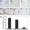
Targeted aV integrin ligand adeno-associated virus phage vector-expressing tumor necrosis factor-α (RGD-A-TNF) induces vascular damage of tumor vessels in vivo. Day 4 tumor sections from animals that received either nontargeted fd-A-TNF (n = 3) (a and d), RGD-A-null (n = 3) (b and e), or RGD-A-TNF (n = 3) (c and f) were stained for CD31, a vascular endothelial cell marker, by immunohistochemical staining. The blood vessels stained brown with anti-CD31-specific antibody (original magnification, ×100 in a-c, ×200 in d-f). The higher magnification highlights an area demonstrating vessel morphology. CD31 staining was quantified and is represented as the percentage area (g). Error bars indicate the standard error of mean.
FIGURE 7.
FIGURE 7.
Inhibition of tumor growth in adeno-associated virus phage vector (AAVP)-treated animals. Nude mice with subcutaneously implanted human melanoma tumors were treated twice (on Day 0 and Day 7) with AAVP systemically through tail vein injection, and tumor volumes were measured at different time points. The tumor volumes (in mm3) were plotted against the different days post-treatment. The animals that were treated with targeted αV integrin ligand (RGD-4C) AAVP-expressing tumor necrosis factor-α (RGD-A-TNF) (circles) had a statistically significant reduction in tumor volumes (P = .048) compared with animals that were treated either with vehicle (diamonds), or nontargeted fd-A-TNF (triangles), or RGD-A-null (squares).

References

    1. Folkman J. Angiogenesis. Annu Rev Med. 2006;57:1–18. - PubMed
    1. ten Hagen TL, Eggermont AM. Solid tumor therapy: manipulation of the vasculature with TNF. Technol Cancer Res Treat. 2003;2:195–203. - PubMed
    1. Scarpati EM, Sadler JE. Regulation of endothelial cell coagulant properties. Modulation of tissue factor, plasminogen activator inhibitors, and thrombomodulin by phorbol 12-myristate 13-acetate and tumor necrosis factor. J Biol Chem. 1989;264:20705–20713. - PubMed
    1. Brett J, Gerlach H, Nawroth P, Steinberg S, Godman G, Stern D. Tumor necrosis factor/cachectin increases permeability of endothelial cell monolayers by a mechanism involving regulatory G proteins. J Exp Med. 1989;169: 1977–1991. - PMC - PubMed
    1. Friedl J, Puhlmann M, Bartlett DL, et al.Induction of permeability across endothelial cell monolayers by tumor necrosis factor (TNF) occurs via a tissue factor-dependent mechanism: relationship between the procoagulant and permeability effects of TNF. Blood. 2002;100:1334–1339. - PubMed

Publication types

Substances