. 2009 Jul;90(1):373-80.
doi: 10.1002/jbm.b.31295.

Host-derived loss of dentin matrix stiffness associated with solubilization of collagen

Affiliations

Host-derived loss of dentin matrix stiffness associated with solubilization of collagen

Marcela R Carrilho et al. J Biomed Mater Res B Appl Biomater. 2009 Jul.

Abstract

Matrix metalloproteinases (MMPs) bound to dentin matrices are activated during adhesive bonding procedures and are thought to contribute to the progressive degradation of resin-dentin bonds over time. The purpose of this study was to evaluate the changes in mechanical, biochemical, and structural properties of demineralized dentin treated with or without chlorhexidine (CHX), a known MMP-inhibitor. After demineralizing dentin beams in EDTA or phosphoric acid (PA), the baseline modulus of elasticity (E) of each beam was measured by three-point flexure. Specimens were pretreated with water (control) or with 2% CHX (experimental) and then incubated in artificial saliva (AS) at 37 degrees C for 4 weeks. The E of each specimen was remeasured weekly and, the media was analyzed for solubilized dentin collagen at first and fourth week of incubation. Some specimens were processed for electron microscopy (TEM) immediately after demineralization and after 4 weeks of incubation. In EDTA and PA-demineralized specimens, the E of the control specimens fell (p < 0.05) after incubation in AS, whereas there were no changes in E of the CHX-pretreated specimens over time. More collagen was solubilized from PA-demineralized controls (p < 0.05) than from EDTA-demineralized matrices after 1 or 4 weeks. Less collagen (p < 0.05) was solubilized from CHX-pretreated specimens demineralized in EDTA compared with PA. TEM examination of control beams revealed that prolonged demineralization of dentin in 10% PA (12 h) did not denature the collagen fibrils.


Figures

Figure 1
Figure 1
Changes in the modulus of elasticity of completely demineralized normal human coronal dentin beams over 4 weeks of incubation. A. 0.5 M EDTA (pH 7.4) demineralized beams. AS - control beams preincubated in water and then incubated in artificial saliva (AS). 2% CHX - experimental beams pretreated with 2% chlorhexidine digluconate and then incubated in AS. B. 10% phosphoric acid (PA) demineralized beams. Abbreviations are the same as in A. Height of bars signifies the mean value, the brackets ± SD, n = 10 in each subgroup. Bars identified by different lower case letters are significantly different (p<0.05).
Figure 1
Figure 1
Changes in the modulus of elasticity of completely demineralized normal human coronal dentin beams over 4 weeks of incubation. A. 0.5 M EDTA (pH 7.4) demineralized beams. AS - control beams preincubated in water and then incubated in artificial saliva (AS). 2% CHX - experimental beams pretreated with 2% chlorhexidine digluconate and then incubated in AS. B. 10% phosphoric acid (PA) demineralized beams. Abbreviations are the same as in A. Height of bars signifies the mean value, the brackets ± SD, n = 10 in each subgroup. Bars identified by different lower case letters are significantly different (p<0.05).
Figure 2
Figure 2
Changes in the modulus of elasticity of completely demineralized normal human coronal dentin beams for 4 weeks of incubation. A. Same data as Fig. 1 but grouped to compare EDTA- vs. PA-demineralized specimens at each time-point in the control (AS) group. B. Same data as in Fig. 1 but grouped to compare EDTA- vs. PA-demineralized specimens in the experimental (2% CHX pretreatment group). Symbols have same meaning as in Fig. 1.
Figure 2
Figure 2
Changes in the modulus of elasticity of completely demineralized normal human coronal dentin beams for 4 weeks of incubation. A. Same data as Fig. 1 but grouped to compare EDTA- vs. PA-demineralized specimens at each time-point in the control (AS) group. B. Same data as in Fig. 1 but grouped to compare EDTA- vs. PA-demineralized specimens in the experimental (2% CHX pretreatment group). Symbols have same meaning as in Fig. 1.
Figure 3
Figure 3
Percent of the total collagen that was solubilized from dentin beams after 1 and 4 weeks of incubation. A. 0.5 EDTA-demineralized beams. The media from both groups was removed at 1 and 4 weeks, hydrolyzed in HCl and analyzed for hydroxyproline, that in turn, premixed calculation of solubilized collagen. More collagen was solubilized after 4 weeks than after 1 week in both control (AS) and experimental (CHX) specimens. Symbols without SD brackets indicates that ± 1 SD was smaller than the size of the symbol. Points identified by different lower case letters are significantly different (p<0.05). N = 10 in all subgroups. B. Specimens completely demineralized in 10% phosphoric acid. Note that significantly more collagen was solubilized at 1 week compared to 4 weeks in both groups but especially in the control (AS) group. C. Same data as in Fig. 3A and B but grouped to permit comparison of the two types of demineralization (EDTA vs. PA) in the control (AS) group at 1 and 4 weeks. D. Data grouped to permit comparison of the two types of demineralization (EDTA vs. PA) in the experimental (CHX) group at 1 and 4 weeks.
Figure 4
Figure 4
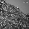
Transmission electron micrographs of demineralized dentin beams. A. A EDTA-demineralized beam immediately after 6 days of demineralization at 25°C. Notice the morphological integrity of collagen fibrils. The granular material on the surface (arrow) may represent residual smear layer material. B. A PA-demineralized beam immediately after demineralization. Although there was a thin (0.5 μm) zone of surface degradation with defibrillation of collagen fibrils, the collagen fibrils below the surface maintained their cross-banding and heavy metal uptake.
Figure 4
Figure 4
Transmission electron micrographs of demineralized dentin beams. A. A EDTA-demineralized beam immediately after 6 days of demineralization at 25°C. Notice the morphological integrity of collagen fibrils. The granular material on the surface (arrow) may represent residual smear layer material. B. A PA-demineralized beam immediately after demineralization. Although there was a thin (0.5 μm) zone of surface degradation with defibrillation of collagen fibrils, the collagen fibrils below the surface maintained their cross-banding and heavy metal uptake.

References

    1. Marshall GW, Jr, Marshall SJ, Kinney JH, Balooch M. The dentin substrate: structure and properties related to bonding. J Dent. 1997;25(6):441–58. - PubMed
    1. Kinney JHJ, Pople A, Marshall GW, Marshall SJ. Collagen orientation and crystallite size in human dentin: a small angle X-ray scattering study. Calcif Tissue Int. 2001;69(1):31–7. - PubMed
    1. Miguez PA, Pereira PN, Atsawasuwan P, Yamauchi M. Collagen cross-linking and ultimate tensile strength in dentin. J Dent Res. 2004;83(10):807–10. - PubMed
    1. Nishitani Y, Yoshiyama M, Tay FR, Wadgaonkar B, Waller J, Agee K, Pashley DH. Tensile strength of mineralized/demineralized human normal and carious dentin. J Dent Res. 2005;84(11):1075–78. - PMC - PubMed
    1. Schlueter RJ, Veis A. The macromolecular organization of dentin matrix collagen. I. Periodate degradation and carbohydrate cross-linking. Biochemistry. 1964;3:1657–65. - PubMed

Publication types