Review
. 2008;10 Suppl 1(Suppl 1):S3.
doi: 10.1186/bcr2126. Epub 2008 Dec 9.

Molecular imaging as a tool for translating breast cancer science

Affiliations
Review

Molecular imaging as a tool for translating breast cancer science

David A Mankoff. Breast Cancer Res. 2008.

Abstract

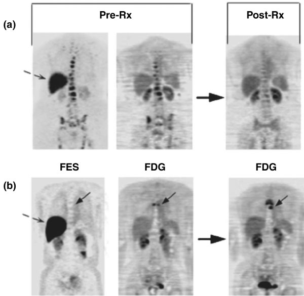
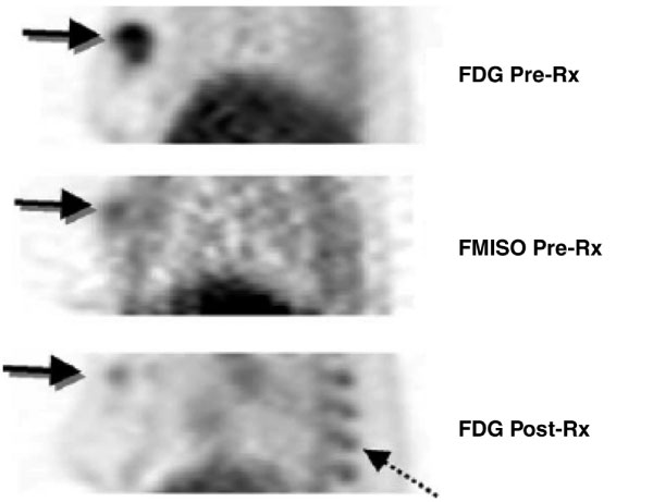
The ability to measure biochemical and molecular processes underlies progress in breast cancer biology and treatment. These assays have traditionally been performed by analysis of cell culture or tissue samples. More recently, functional and molecular imaging has allowed the in vivo assay of biochemistry and molecular biology, which is highly complementary to tissue-based assays. This review briefly describes different imaging modalities used in molecular imaging and then reviews applications of molecular imaging to breast cancer, with a focus on translational work. It includes sections describing work in functional and physiological tumor imaging, imaging gene product expression, imaging the tumor microenvironment, reporter gene imaging, and cell labeling. Work in both animal models and human is discussed with an eye towards studies that have relevance to breast cancer treatment in patients.


Figures

Figure 1
Figure 1
Imaging examples: pre-treatment (Pre-Rx) 16 alpha-[18F]-fluoro-17 beta-estradiol positron emission tomography (FES PET; left) and 18F-fluorodeoxyglucose (FDG) PET (middle) scans and follow-up FDG PET post-therapy (Post-Rx; right) are shown. Dashed arrows show normal liver FES uptake. (a) This patient has bone metastasis with robust FES and FDG uptake, and had response at 3 months. (b) This patient has bone metastasis (solid arrow) without FES but with FDG uptake; progressive disease at 6 months. (Reproduced from [94].)
Figure 2
Figure 2
Breast tumor hypoxia as a predictor of drug resistance. A patient with a large, right breast tumor underwent 18F-fluorodeoxyglucose (FDG) and 18F-fluoromisonidazole (FMISO) positron emission tomography (PET) pre-therapy (Pre-Rx; top and middle) and after approximately ten weeks of chemotherapy (Post-Rx; bottom). Images are thick sagittal images, similar to medial-lateral oblique (MLO) mammography views. The pre-therapy FDG study showed uniformly high FDG uptake throughout the tumor. FMISO PET showed uptake suggestive of tumor hypoxia, but only close to the center of the tumor (arrow). Post-therapy images show a dramatic reduction in the extent and intensity of FDG uptake with residual activity in the part of the tumor that had FMISO uptake pre-therapy. Residual viable tumor was found at surgery. Marrow uptake of FDG was also seen post-therapy (dashed arrow) because of granulocyte colony-stimulating factor administered for marrow support as part of the treatment. (Reproduced from [158].)

Similar articles

Cited by

References

    1. Hutter H. Fluorescent reporter methods. Methods Mol Biol. 2006;351:155–173. - PubMed
    1. Blasberg RG. Imaging update: new windows, new views. Clin Cancer Res. 2007;13:3444–3448. - PubMed
    1. Mankoff DA. A definition of molecular imaging. J Nucl Med. 2007;48:18N. 21N. - PubMed
    1. Mankoff DA, O'Sullivan F, Barlow WE, Krohn KA. Molecular imaging research in the outcomes era: measuring outcomes for individualized cancer therapy. Acad Radiol. 2007;14:398–405. - PMC - PubMed
    1. Benard F, Turcotte E. Imaging in breast cancer: single-photon computed tomography and positron-emission tomography. Breast Cancer Res. 2005;7:153–162. - PMC - PubMed