. 2008 Dec 17:8:373.
doi: 10.1186/1471-2407-8-373.

Desmoglein 2 is a substrate of kallikrein 7 in pancreatic cancer

Affiliations

Desmoglein 2 is a substrate of kallikrein 7 in pancreatic cancer

Vishnu C Ramani et al. BMC Cancer. .

Abstract

Background: In a previous report we have demonstrated that the chymotryptic-like serine protease kallikrein 7 (KLK7/hK7) is overexpressed in pancreatic cancer. In normal skin, hK7 is thought to participate in skin desquamation by contributing in the degradation of desmosomal components, such as desmogleins. Thus, the ability of hK7 to degrade desmogleins was assessed and the effect of hK7 expression on desmoglein 2 was examined in cultured pancreatic cancer cells.

Methods: The expression of Dsg1, Dsg2, and Dsg3 in pancreatic tissues was examined by immunohistochemistry and their expression in two pancreatic cancer cell lines, BxPC-3 and Panc-1, was determined by western blot analysis. The ability of hK7 to degrade Dsg1 and Dsg2 was investigated using in vitro degradation assays. BxPC-3 cells stably transfected to overexpress hK7 were used to examine the effect of hK7 on cell-surface resident Dsg2.

Results: The levels of immunoreactive Dsg1 and Dsg2 were reduced in pancreatic adenocarcinomas compared with both normal pancreatic and chronic pancreatitis tissues. Among the desmosomal proteins examined, Dsg2 exhibited robust expression on the surface of BxPC-3 cells. When hK7 was overexpressed in this cell line, there was a significant increase in the amount of soluble Dsg2 released into the culture medium compared with vector-transfected control cells.

Conclusion: A reduction in the amount of the cell adhesion components Dsg1 and Dsg2 in pancreatic tumors suggests that loss of these desmosomal proteins may play a role in pancreatic cancer invasion. Using in vitro degradation assays, both Dsg1 and Dsg2 could be readily proteolyzed by hK7, which is overexpressed in pancreatic adenocarcinomas. The enforced expression of hK7 in BxPC-3 cells that express significant amounts of Dsg2 resulted in a marked increase in the shedding of soluble Dsg2, which is consistent with the notion that aberrant expression of hK7 in pancreatic tumors may result in diminished cell-cell adhesion and facilitate tumor cell invasion.


Figures
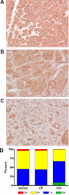
Figure 1
Figure 1
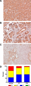
Desmoglein 1 is present in lower levels in pancreatic cancer compared to normal and chronic pancreatitis tissue. Representative pancreatic tissue sections stained for Dsg1 showed high membrane staining in (A) normal pancreas, (B) chronic pancreatitis (CP), and (C) weaker staining in pancreatic adenocarcinoma (PaC) samples. Original magnification x400. (D) Staining intensity of the cell membranes was quantitated (see Methods), categorized into intensity ranges from 0+ to 3+, and the average percentage of cells in each group was determined and represented in a stacked graph for each tissue type (n = 6).
Figure 2
Figure 2
Desmoglein 2 staining is lower in pancreatic cancer cell membranes compared to normal and chronic pancreatitis tissue. Representative pancreatic tissue sections stained for Dsg2 showed high levels of membrane staining in (A) normal pancreas and (B) chronic pancreatitis (CP) samples. Membrane staining was much weaker in (C) pancreatic adenocarcinoma (PaC) samples. Original magnification x400. (D) Staining intensity of the cell membranes was quantitated (see Methods), categorized into intensity ranges from 0+ to 3+, and the average percentage of cells in each group was determined and represented in a stacked graph for each tissue type (n = 6).
Figure 3
Figure 3
Desmoglein 1 and 2 are substrates for hK7 in vitro. Thermolysin-activated hK7 was incubated with recombinant Dsg1 (A) or Dsg2 (B) for the indicated times in the absence (lanes 1–4) or presence (lanes 5–8) of the hK7 inhibitor chymostatin. Reaction products were separated by SDS-PAGE and visualized by western blot using either anti-Dsg1 or anti-Dsg2 antibodies. Sizes of protein markers are indicated on the left.
Figure 4
Figure 4
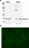
Expression of desmogleins in human pancreatic adenocarcinoma cell lines. (A) Equal amounts of total protein from whole cell lysates of human pancreatic cancer cell lines BxPC-3 (lanes 1 and 3) and Panc-1 (lanes 2 and 4) were separated by SDS-PAGE and visualized by western blot using anti-Dsg1 and anti-Dsg2 antibodies, as indicated. GAPDH levels were monitored as a loading control (arrowhead). Sizes of protein markers are indicated on the left. Appreciable levels of Dsg2 were detected in BxPC-3 cells, but not in Panc-1 cells. Dsg1 was not detected in either of the cell lines. (B) Immunocytochemistry of Dsg2 in BxPC-3 cells using a FITC-conjugated secondary antibody showed specific cell membrane localization. Original magnification x400.
Figure 5
Figure 5
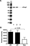
Levels of soluble Dsg2 in conditioned medium of KLK7-expressing BxPC-3 cells are higher than in vector-transfected control cells. (A) Levels of soluble Dsg2 (sDsg2) in concentrated conditioned medium from hK7-expressing clones BxPC-3/hK7 clone#3 (lane 1) and clone#7 (lane 2) and vector-transfected clone BxPC-3/Vec (lane 3) were determined by western blot. Sizes of protein markers are indicated on the left. (B) The relative amount of the 80-kDa sDsg2 immunoreactive product was determined for KLK7-transfected (solid bars) and vector-transfected (open bar) BxPC-3 cells by densitometric analysis using Quantity One software and the average relative intensity is depicted. Vertical bars represent SEM (n = 3).

Similar articles

Cited by

References

    1. Hanahan D, Weinberg RA. The hallmarks of cancer. Cell. 2000;100:57–70. - PubMed
    1. Zetter BR. Adhesion molecules in tumor metastasis. Semin Cancer Biol. 1993;4:219–229. - PubMed
    1. Green KJ, Gaudry CA. Are desmosomes more than tethers for intermediate filaments? Nat Rev Mol Cell Biol. 2000;1:208–216. - PubMed
    1. Kowalczyk AP, Bornslaeger EA, Norvell SM, Palka HL, Green KJ. Desmosomes: intercellular adhesive junctions specialized for attachment of intermediate filaments. Int Rev Cytol. 1999;185:237–302. - PubMed
    1. Chidgey M, Dawson C. Desmosomes: a role in cancer? Br J Cancer. 2007;96:1783–1787. - PMC - PubMed

Publication types