. 2008 Dec 18:8:375.
doi: 10.1186/1471-2407-8-375.

Increased p38-MAPK is responsible for chemotherapy resistance in human gastric cancer cells

Affiliations

Increased p38-MAPK is responsible for chemotherapy resistance in human gastric cancer cells

Xianling Guo et al. BMC Cancer. .

Abstract

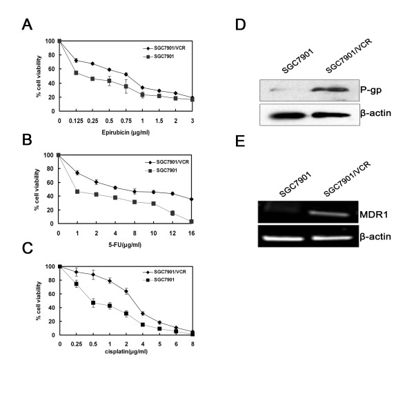
Background: Chemoresistance is one of the main obstacles to successful cancer therapy and is frequently associated with Multidrug resistance (MDR). Many different mechanisms have been suggested to explain the development of an MDR phenotype in cancer cells. One of the most studied mechanisms is the overexpression of P-glycoprotein (P-gp), which is a product of the MDR1 gene. Tumor cells often acquire the drug-resistance phenotype due to upregulation of the MDR1 gene. Overexpression of MDR1 gene has often been reported in primary gastric adenocarcinoma.

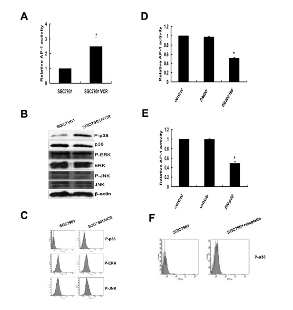
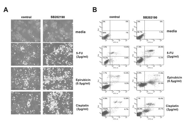
Methods: This study investigated the role of p38-MAPK signal pathway in vincristine-resistant SGC7901/VCR cells. P-gp and MDR1 RNA were detected by Western blot analysis and RT-PCR amplification. Mitgen-activated protein kinases and function of P-gp were demonstrated by Western blot and FACS Aria cytometer analysis. Ap-1 activity and cell apoptosis were detected by Dual-Luciferase Reporter Assay and annexin V-PI dual staining.

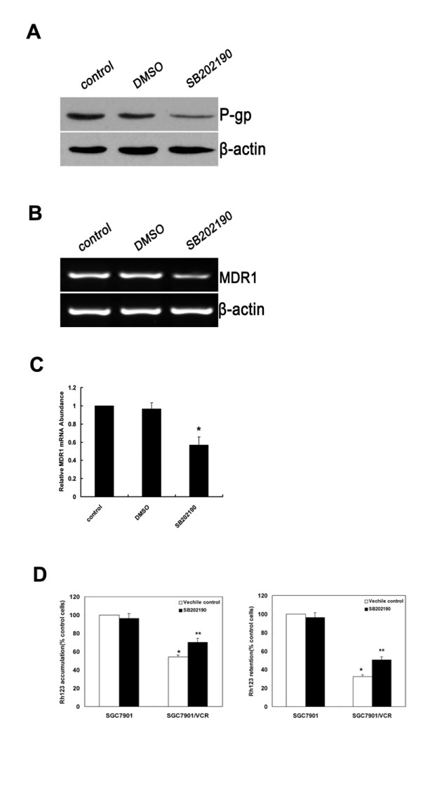
Results: The vincristine-resistant SGC7901/VCR cells with increased expression of the multidrug-resistance 1 (MDR1) gene were resistant to P-gp-related drug and P-gp-unrelated drugs. Constitutive increases of phosphorylated p38-MAPK and AP-1 activities were also found in the drug-resistant cells. Inhibition of p38-MAPK by SB202190 reduced activator protein-1 (AP-1) activity and MDR1 expression levels and increased the sensitivity of SGC7901/VCR cells to chemotherapy.

Conclusion: Activation of the p38-MAPK pathway might be responsible for the modulation of P-glycoprotein-mediated and P-glycoprotein-unmediated multidrug resistance in the SGC7901/VCR cell line.


Figures

Figure 1
Figure 1
SGC7901/VCR cells that overexpress MDR1 are more resistant to chemotherapy than SGC7901 cells. (A-C) After treatment with different concentrations of 5-FU, cisplatin or epirubicin for 72 hours, cell viability was determined using the MTT assay. The viability of the untreated cells was taken as 100%. Points, mean of three independent experiments; bars, SD. Significant differences are indicated by asterisks. *, P < 0.05. (D) SGC7901/VCR and SGC7901 cells were harvested to prepare cell lysates. The lysates were subjected to SDS-PAGE and blotted with anti-P-gp antibody. (E) RT-PCR assays were performed to detect MDR1 mRNA. A representative example of an experiment that was repeated three times is shown.
Figure 2
Figure 2
Activation of p38 MAPK/AP-1 pathway in SGC7901/VCR cells. (A) SGC7901/VCR and SGC7901 cells were transfected with AP-1 luciferase reporter plasmids. After 24 hours, the cells were collected and luciferase assays were performed. The luciferase activity results in SGC7901 cells were normalized to 1.0. Error bars indicate standard deviations. The values shown represent the means of at least three separate experiments. Significant differences are indicated by asterisks. *, P < 0.05. (B) Levels of phosphorylated and non-phosphroylated MAP kinases (p38-MAPK, ERKs and JNKs) in SGC7901/VCR and SGC7901 cells were detected using Western-blot analyses. A representative example of an experiment that was repeated three times is shown. (C) FACS analysis of p38-MAPK, ERK and JNK phosphorylation. A representative example of an experiment that was repeated three times is shown. (D-E) Analysis of AP-1 activity using the luciferase assay in SGC7901/VCR cells treated or untreated with SB202190 (10 μM), or cotransfected with the DN-p38 plasmid for 24 hours. The luciferase activity in control samples was normalized to 1.0. Error bars indicate standard deviations. The values shown represent the means of at least three separate experiments. Significant differences are indicated by asterisks. *, P < 0.05. (F) SGC7901 cells were incubated with cisplatin (2 μg/ml) for 24 h, and then cells were collected. FACS analysis was used to detect p38-MAPK phosphorylation.
Figure 3
Figure 3
Inhibition of p38-MAPK impairs MDR1 expression and function of P-gp in SGC7901/VCR cells. SGC7901/VCR cells were treated with DMSO or SB202190 (10 μM). (A) Protein levels of P-gp were detected by Western-blot analysis. A representative example of an experiment that was repeated three times is shown. (B) SGC7901/VCR cells were treated with DMSO and SB202190. Expression of MDR1 mRNA was assessed by RT-PCR. β-actin mRNA levels were measured as positive internal controls. (C) The MDR1 mRNA expression levels were normalized to those of β-actin and are the means ± SD of at least three independent experiments. Significant differences are indicated by asterisks. *, P < 0.05. (D)Effects of SB202190 on Rh123 accumulation (left) and retention (right) in SGC7901 and SGC7901/VCR cells. (left) Cells treated with SB202190 (10 μM) or vehicle control (0.1% DMSO). Rh123 (1 μg/mL) was added, and the cells were incubated for 120 min. (right) Cells were incubated with Rh123 for 120 min, washed, and resuspended in medium with SB202190 (10 μM)or vehicle control (0.1% DMSO) for 120 min. Rh123 fluorescence was measured using FAC scan. Means ± SD from three independent experiments. *P < 0.05 vs SGC7901 cells. **P < 0.05 vs vehicle control.
Figure 4
Figure 4
Inhibition of p38 MAPK increases SGC7901/VCR to chemotherapy. SGC7901/VCR cells were treated with indicated concentrations of cisplatin, 5-FU or epirubicin, with or without SB202190 (10 μM), for 24 hours. (A) The morphology of cells was observed, and photographs were taken under the microscope. (B) Flow cytometry analysis was performed after staining with Annexin V/PI. Compared with the control cells (without SB202190), there were significantly more apoptotic cells in the SB230920 treatment group. A representative example of an experiment that was repeated three times is shown.

Similar articles

Cited by

References

    1. Gottesman MM, Fojo T, Bates SE. Multidrug resistance in cancer: role of ATP-dependent transporters. Nat Rev Cancer. 2002;2:48–58. doi: 10.1038/nrc706. - DOI - PubMed
    1. Endicott JA, Ling V. The biochemistry of P-glycoprotein-mediated multidrug resistance. Annu Rev Biochem. 1989;58:137–171. doi: 10.1146/annurev.bi.58.070189.001033. - DOI - PubMed
    1. Ambudkar SV, Dey S, Hrycyna CA, Ramachandra M, Pastan I, Gottesman MM. Biochemical, cellular, and pharmacological aspects of the multidrug transporter. Annu Rev Pharmacol Toxicol. 1999;39:361–398. doi: 10.1146/annurev.pharmtox.39.1.361. - DOI - PubMed
    1. Hait WN, Yang JM. Clinical management of recurrent breast cancer: development of multidrug resistance (MDR) and strategies to circumvent it. Semin Oncol. 2005;32:S16–21. doi: 10.1053/j.seminoncol.2005.09.011. - DOI - PubMed
    1. Mickisch G, Fajta S, Bier H, Tschada R, Alken P. Cross-resistance patterns related to glutathione metabolism in primary human renal cell carcinoma. Urol Res. 1991;19:99–103. doi: 10.1007/BF00368184. - DOI - PubMed

Publication types

MeSH terms

Substances