

Protein scaffolds in MAP kinase signalling
- PMID: 19091303
- PMCID: PMC2668224
- DOI: 10.1016/j.cellsig.2008.11.013
Protein scaffolds in MAP kinase signalling
Abstract
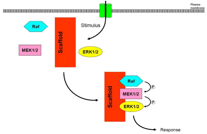
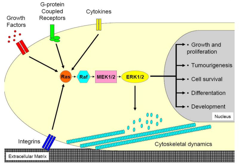
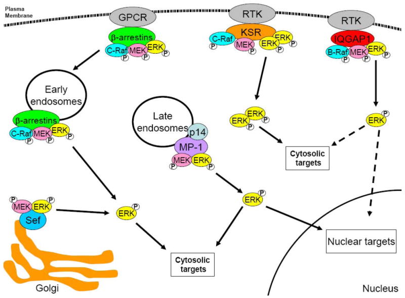
The mitogen-activated protein kinase (MAPK) pathway allows cells to interpret external signals and respond in an appropriate way. Diverse cellular functions, ranging from differentiation and proliferation to migration and inflammation, are regulated by MAPK signalling. Therefore, cells have developed mechanisms by which this single pathway modulates numerous cellular responses from a wide range of activating factors. This specificity is achieved by several mechanisms, including temporal and spatial control of MAPK signalling components. Key to this control are protein scaffolds, which are multidomain proteins that interact with components of the MAPK cascade in order to assemble signalling complexes. Studies conducted on different scaffolds, in different biological systems, have shown that scaffolds exert substantial control over MAPK signalling, influencing the signal intensity, time course and, importantly, the cellular responses. Protein scaffolds, therefore, are integral elements to the modulation of the MAPK network in fundamental physiological processes.
Figures
References
-
- Pearson G, Robinson F, Beers Gibson T, Xu BE, Karandikar M, Berman K, Cobb MH. Endocr Rev. 2001;22(2):153–183. - PubMed
-
- Murphy LO, Blenis J. Trends Biochem Sci. 2006;31(5):268–275. - PubMed
-
- Mor A, Philips MR. Annu Rev Immunol. 2006;24:771–800. - PubMed
-
- Wellbrock C, Karasarides M, Marais R. Nat Rev Mol Cell Biol. 2004;5(11):875–885. - PubMed
Publication types
MeSH terms
Substances
Grants and funding
LinkOut - more resources
Full Text Sources
Other Literature Sources
Molecular Biology Databases
