

The role of trauma-related distractors on neural systems for working memory and emotion processing in posttraumatic stress disorder
- PMID: 19091328
- PMCID: PMC2684984
- DOI: 10.1016/j.jpsychires.2008.10.014
The role of trauma-related distractors on neural systems for working memory and emotion processing in posttraumatic stress disorder
Abstract
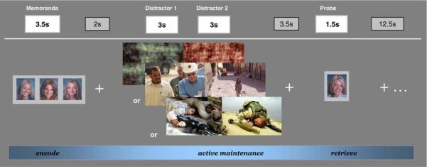
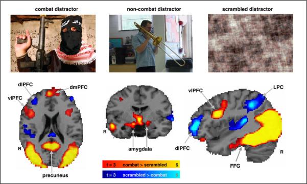
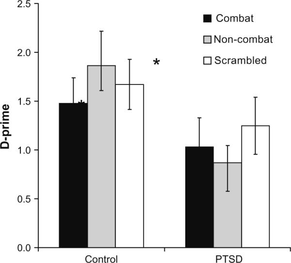
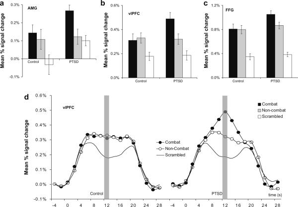
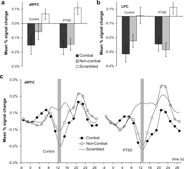
The relevance of emotional stimuli to threat and survival confers a privileged role in their processing. In PTSD, the ability of trauma-related information to divert attention is especially pronounced. Information unrelated to the trauma may also be highly distracting when it shares perceptual features with trauma material. Our goal was to study how trauma-related environmental cues modulate working memory networks in PTSD. We examined neural activity in participants performing a visual working memory task while distracted by task-irrelevant trauma and non-trauma material. Recent post-9/11 veterans were divided into a PTSD group (n=22) and a trauma-exposed control group (n=20) based on the Davidson trauma scale. Using fMRI, we measured hemodynamic change in response to emotional (trauma-related) and neutral distraction presented during the active maintenance period of a delayed-response working memory task. The goal was to examine differences in functional networks associated with working memory (dorsolateral prefrontal cortex and lateral parietal cortex) and emotion processing (amygdala, ventrolateral prefrontal cortex, and fusiform gyrus). The PTSD group showed markedly different neural activity compared to the trauma-exposed control group in response to task-irrelevant visual distractors. Enhanced activity in ventral emotion processing regions was associated with trauma distractors in the PTSD group, whereas activity in brain regions associated with working memory and attention regions was disrupted by distractor stimuli independent of trauma content. Neural evidence for the impact of distraction on working memory is consistent with PTSD symptoms of hypervigilance and general distractibility during goal-directed cognitive processing.
Figures
References
-
- Beck AT, Steer RA, Garbin MG. Psychometric properties of the beck depression inventory: twenty-five years of evaluation. Clinical Psychology Review. 1988;8(1):77–100.
-
- Bremner J, Vermetter E, Vythilingam M, Afzal N, Schmahl C, Bernet Elzinga, et al. Neural correlates of the classic color and emotional stroop in women with abuse-related posttraumatic stress disorder. Biological Psychiatry. 2004;55:612–20. - PubMed
-
- Bryant RA, Felmingham KL, Kemp AH, Barton M, Peduto AS, Rennie C, et al. Neural networks of information processing in posttraumatic stress disorder: a functional magnetic resonance imaging study. Biological Psychiatry. 2005;58(2):111–8. - PubMed
-
- Clark CR, McFarlane AC, Morris P, Weber DL, Sonkkilla C, Shaw M, et al. Cerebral function in posttraumatic stress disorder during verbal working memory updating: a positron emission tomography study. Biological Psychiatry. 2003;53:474–81. - PubMed
-
- Corbetta M, Shulman GL. Control of goal-directed and stimulus-driven attention in the brain. Nature Reviews Neuroscience. 2002;3(3):201–15. - PubMed
Publication types
MeSH terms
Grants and funding
LinkOut - more resources
Full Text Sources
Medical
