. 2009 Feb 20;284(8):5056-66.
doi: 10.1074/jbc.M808128200. Epub 2008 Dec 17.

Slc2a5 (Glut5) is essential for the absorption of fructose in the intestine and generation of fructose-induced hypertension

Affiliations

Slc2a5 (Glut5) is essential for the absorption of fructose in the intestine and generation of fructose-induced hypertension

Sharon Barone et al. J Biol Chem. .

Abstract

The identity of the transporter responsible for fructose absorption in the intestine in vivo and its potential role in fructose-induced hypertension remain speculative. Here we demonstrate that Glut5 (Slc2a5) deletion reduced fructose absorption by approximately 75% in the jejunum and decreased the concentration of serum fructose by approximately 90% relative to wild-type mice on increased dietary fructose. When fed a control (60% starch) diet, Glut5(-/-) mice had normal blood pressure and displayed normal weight gain. However, whereas Glut5(+/+) mice showed enhanced salt absorption in their jejuna in response to luminal fructose and developed systemic hypertension when fed a high fructose (60% fructose) diet for 14 weeks, Glut5(-/-) mice did not display fructose-stimulated salt absorption in their jejuna, and they experienced a significant impairment of nutrient absorption in their intestine with accompanying hypotension as early as 3-5 days after the start of a high fructose diet. Examination of the intestinal tract of Glut5(-/-) mice fed a high fructose diet revealed massive dilatation of the caecum and colon, consistent with severe malabsorption, along with a unique adaptive up-regulation of ion transporters. In contrast to the malabsorption of fructose, Glut5(-/-) mice did not exhibit an absorption defect when fed a high glucose (60% glucose) diet. We conclude that Glut5 is essential for the absorption of fructose in the intestine and plays a fundamental role in the generation of fructose-induced hypertension. Deletion of Glut5 results in a serious nutrient-absorptive defect and volume depletion only when the animals are fed a high fructose diet and is associated with compensatory adaptive up-regulation of ion-absorbing transporters in the colon.


Figures

FIGURE 1.
FIGURE 1.
Expression of Glut5 in small intestine and kidney of Glut+/+ and Glut5-/- mice. a, Northern hybridization. Expression of Glut5 was completely absent in small intestine and kidney of Glut5-/- mice. b, immunofluorescence labeling. Glut5 labeling was completely absent in jejunum of Glut5-/- mice. c, transient expression of Glut5 in cultured cells: [14C]fructose or [14C]glucose uptake. COS-7 cells were transiently transfected with the Glut5 cDNA. The uptake of 50 μmol of [14C]fructose or [14C]glucose was measured in cultured cells at 10 min (left and middle panels). d, [14C]fructose or [14C]glucose influx in luminal membrane vesicles from jejunum. The membrane vesicles were preincubated in a solution that contained 10 mm HEPES-Tris, and 135 mm NaCl, pH 7.5. The uptake started by adding 10 μl of brush border membrane vesicles to 30 μl of medium containing 10 mm HEPES-Tris, 135 mm NaCl, at pH 7.5 and 50 μmol of unlabeled fructose or glucose and [14C]fructose or [14C]glucose tracer. The uptake experiments were assayed at room temperature in triplicate by the rapid filtration technique.
FIGURE 2.
FIGURE 2.
Role of Glut5 in dietary fructose absorption. a, body weight in Glut5+/+ and Glut5-/- mice fed a high fructose or control diet. Unlike Glut5+/+ mice, Glut5-/- mice experienced significant weight loss on the high fructose diet. b, blood fructose concentration in Glut5+/+ and Glut5-/- mice fed a high fructose or control diet. Unlike Glut5+/+ mice, fructose concentration did not increase in Glut5-/- mice on the high fructose diet.
FIGURE 3.
FIGURE 3.
Glut5 deletion causes malabsorption in mice fed a high fructose diet. a, colon dilatation in mice on high fructose. Top right panel shows Glut5-/- mice on the high fructose diet had distended caecum and proximal colon. Glut5+/+ mice on high fructose (top left) and Glut5+/+ and Glut5-/- mice on control diet (bottom panels) display normal bowel morphology. b, gross anatomy of intestinal tract in mice on high fructose. All segments of the intestinal tract showed enlargement, and the caecum and colon showed distension in Glut5-/- mice on the high fructose diet due to increased amount of luminal contents resulting from malabsorption. c, cecal contents. The content of caecum in Glut5-/- mice is significantly increased (∼10-fold) as compared with Glut5+/+ mice on the high fructose diet. d, intestinal segment size. Mean lengths (small intestine and colon) or area (caecum) of intestinal segments of Glut5+/+ and Glut5-/- mice on control or high fructose diet.
FIGURE 4.
FIGURE 4.
Regulation of Glut2 and Sglt1 in the small intestine by high fructose or high glucose diet in Glut5 null mice. a, Northern hybridization of Glut2 and Sglt1 in the small intestine of Glut5+/+ and Glut5-/- mice on control or high fructose diet. Glut2 expression increased significantly (∼80%) in jejunum of Glut5+/+ mice on the high fructose diet but did not show any up-regulation in Glut5-/- mice on control or high fructose diet. Sglt1 expression increased by 130% in Glut5+/+ but by only ∼25% in Glut5-/- mice on the high fructose diet relative to control diet. b, expression of Glut2 and Sglt1 by quantitative PCR in the small intestine of Glut5+/+ and Glut5-/- mice on control or high fructose diet. Glut2 expression increased by ∼70% in jejunum of Glut5+/+ mice on the high fructose diet but showed no increase in jejunum of Glut5-/- mice on the high fructose diet. Sglt1 expression was enhanced significantly (∼140%) in jejunum of Glut5+/+ mice on the high fructose diet but showed only a mild up-regulation (∼30%) in jejunum of Glut5-/- mice on the high fructose diet. c, immunofluorescence labeling of Glut2 in jejuna of Glut5+/+ and Glut5-/- mice on control or high fructose diet. Glut2 was localized on the basolateral membrane of intestinal villi in Glut5+/+ mice on control diet but was recruited to the apical membrane domain in Glut5+/+ mice on the high fructose diet. Glut2 was detected on both the apical and basolateral membrane in Glut5-/- mice on control or high fructose diet. d, Northern hybridization of Glut2 and Sglt1 in the small intestine of Glut5+/+ and Glut5-/- mice on control or high glucose diet. Glut2 and Sglt1 expression increased significantly and comparably in jejunum of Glut5+/+ and Glut5-/- mice on high glucose diet. e, expression of Glut2 and Sglt1 by quantitative PCR in the small intestine of Glut5+/+ and Glut5-/- mice on control or high glucose diet. Glut2 and Sglt1 expression enhanced by ∼130 and 240%, respectively, in jejunum of Glut5+/+ and by ∼110 and ∼190%, respectively, in jejunum of Glut5-/- mice on high glucose diet.
FIGURE 5.
FIGURE 5.
Effect of luminal fructose on salt absorption in the jejunum and fructose-induced hypertension. a, basal and fructose-stimulated jejunal fluid absorption in Glut5+/+ and Glut5-/- mice. 40 mm fructose in the perfusate elicited an increase of ∼40% in fluid absorption in Glut5+/+ mice jejunum but had no effect in Glut5-/- mice. #, p < 0.01 as compared with NaCl control in Glut5+/+ mice. b, Glut5 up-regulation in jejunum of Glut5+/+ mice fed a high fructose diet for 14 weeks. The expression of Glut5 increased by ∼8-fold in jejunum of Glut5+/+ mice fed a high fructose diet. c, fructose-induced hypertension in Glut5+/+ mice. Systolic blood pressure increased significantly in Glut5+/+ mice on increased dietary fructose intake for 14 weeks. d, effect of high fructose diet on blood pressure in Glut5-/- mice. The systolic blood pressure in Glut5-/- mice was significantly decreased after 5 days on the high fructose diet. Glut5-/- mice developed hypovolemic shock on the high fructose diet for 7–10 days and died.
FIGURE 6.
FIGURE 6.
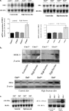
Adaptive regulation of ion transporters in the proximal and distal colon in Glut5-/- mice on increased dietary fructose intake. a, Northern hybridization of NHE3 and DRA in proximal colon. The expression of NHE3 and DRA in the proximal colon increased in Glut5-/- mice on the high fructose diet. Densitometric analysis of NHE3 and DRA expression is shown relative to 28 S rRNA. b, immunoblot analysis of NHE3 and DRA in proximal colon. The expression of NHE3 and DRA increased by ∼320 and 210% in the proximal colon of Glut5-/- mice on the high fructose diet relative to Glut5+/+ mice on the same diet. β-Actin blots are shown as index of protein loading. c, Northern hybridization of colonic H-K ATPase in distal colon. The expression of H-K-ATPase decreased in Glut5-/- mice on high fructose or control diet relative to Glut5+/+ mice (left panel). However, the expression of H-K-ATPase increased in distal colon of DRA null mice (right panel).

References

    1. Hirabayashi, J. (1996) Q. Rev. Biol. 71 365-380 - PubMed
    1. Bollen, M., Keppens, S., and Stalmans, W. (1998) Biochem. J. 336 19-31 - PMC - PubMed
    1. Macdonald, I. A. (1999) Eur. J. Clin. Nutr. 53 Suppl. 1, S101-S106 - PubMed
    1. Rumessen, J. J. (1992) Scand. J. Gastroenterol. 27 819-828 - PubMed
    1. Van den Berghe, G. (1994) Annu. Rev. Nutr. 14 41-58 - PubMed

Publication types

MeSH terms