. 2009 Feb;136(3):495-505.
doi: 10.1242/dev.030007. Epub 2008 Dec 17.

Prox1 maintains muscle structure and growth in the developing heart

Affiliations

Prox1 maintains muscle structure and growth in the developing heart

Catherine A Risebro et al. Development. 2009 Feb.

Erratum in

  • Development. 2009 Feb;136(4):699

Abstract

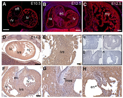
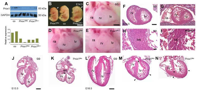
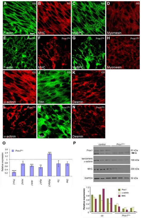
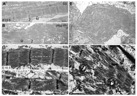
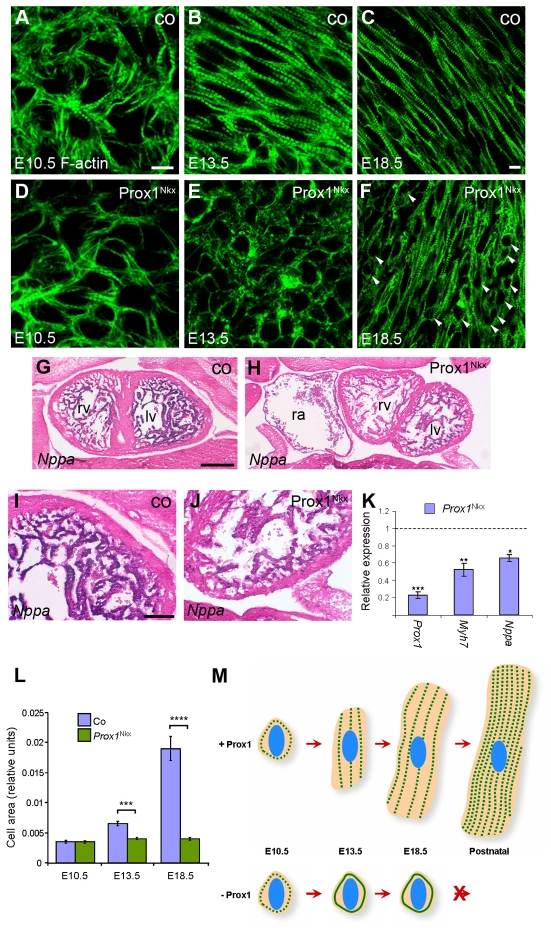
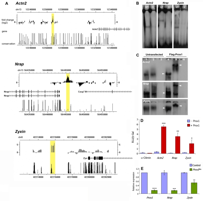
Impaired cardiac muscle growth and aberrant myocyte arrangement underlie congenital heart disease and cardiomyopathy. We show that cardiac-specific inactivation of the murine homeobox transcription factor Prox1 results in the disruption of expression and localisation of sarcomeric proteins, gross myofibril disarray and growth-retarded hearts. Furthermore, we demonstrate that Prox1 is required for direct transcriptional regulation of the genes encoding the structural proteins alpha-actinin, N-RAP and zyxin, which collectively function to maintain an actin-alpha-actinin interaction as the fundamental association of the sarcomere. Aspects of abnormal heart development and the manifestation of a subset of muscular-based disease have previously been attributed to mutations in key structural proteins. Our study reveals an essential requirement for direct transcriptional regulation of sarcomere integrity, in the context of enabling foetal cardiomyocyte hypertrophy, maintenance of contractile function and progression towards inherited or acquired myopathic disease.


Figures

Fig. 1.
Fig. 1.
Prox1 is expressed in the myocardium of the developing mouse heart. (A-C) Immunofluorescence on E10.5 (A) and E12.5 (B,C) frontal sections using an antibody for Prox1. Prox1 is expressed throughout the myocardium of the presumptive left and right ventricles and outflow tract at E10.5 (A) and is localised to both atrial and ventricular myocardium at E12.5 (B,C). (D-H) Immunohistochemistry on E14.5 frontal sections illustrates continued expression of Prox1 throughout the entire myocardium (D), in the interventricular septum (E), the endocardium of the atrioventricular canal endocardial cushions (F,G) and mitral valve leaflets (F,H). Prox1 is absent from cushion mesenchyme (F,G). Panels e-h show the no-primary-antibody controls for the corresponding panels E-H. lv, left ventricle; rv, right ventricle; ivs, interventricular septum; ec, endocardial cushion; en, endocardium; mes, mesenchyme; oft; outflow tract; ra, right atrium; my, myocardium. Scale bars: 50 μm in A,B,D; 25 μm in F; 10 μm in C,E,G,H.
Fig. 2.
Fig. 2.
Cardiac-specific loss of Prox1 perturbs embryonic and heart development. (A) Western blots of E13.5 control (co), Prox1Nkx and Prox1MLC individual isolated mouse heart lysates for Prox1 and Gapdh and (beneath) quantification of protein levels, as normalised to Gapdh, using scanning densitometry. (B-N) Bright-field whole-mount left lateral views of E14.5 Prox1Nkx, Prox1MLC and control (co) littermate embryos (B), and frontal views of hearts in control (C), Prox1Nkx (D) and Prox1MLC (E) embryos. Frontal sections through E14.5 control (F,H) and Prox1Nkx (G,I) embryos, E13.5 isolated control (J) and Prox1Nkx (K) hearts, and E18.5 isolated control (L) and Prox1Nkx (M,N) hearts. Prox1 protein levels are reduced to around a third of control levels in Prox1-conditional hearts (see A). Prox1Nkx mutants are small, with oedema and cranial haemorrhaging, and Prox1MLC mutants reveal extensive oedema (B, arrowheads). Prox1Nkx hearts are hypoplastic with dilation of the right atrium (white dashed lines in C,D; G), and Prox1MLC hearts are hypoplastic with reduced left ventricular expansion (E). Sections through Prox1Nkx hearts reveal myocardial disarray, particularly in the interventricular septum (H,I). By E18.5, Prox1Nkx hearts are rounded in shape and smaller than control hearts, the ventricular wall surface is irregular (arrowheads in M,N) with reduced compaction (black lines in L,M) and there are muscular septal defects (asterisk in N). Also note the membranous ventricular septal defect in Prox1Nkx hearts (arrow, K,M). lv, left ventricle; rv, right ventricle; ra, right atrium; ivs, interventricular septum; ec, endocardial cushion. Scale bars: 5 mm in B; 50 μm in C-G,J,K; 10 μm in H,I; 1 mm in L-N.
Fig. 3.
Fig. 3.
All structural components of the sarcomere are severely disrupted in Prox1-conditional myocardium. (A-N) Confocal sections of immunostained E13.5 whole-mount hearts from control (co; A-D,I-K) and Prox1Nkx (E-H,L-N) mouse embryos. Actin thin filaments and myosin thick filaments lack organisation and are not striated in Prox1-conditional hearts, as visualised by phalloidin staining (green; compare E with A) and immunostaining for sarcomeric myosin heavy chain (MHC) (red; B,F), respectively. Immunostaining for the thick filament component sarcomeric and cardiac myosin binding protein C (MyBP-C) further demonstrates thick filament disorganisation (green; C,G). M-band disruption is demonstrated by immunostaining for myomesin (red; D,H). Z-disc disruption in Prox1Nkx hearts is revealed by immunostaining for sarcomeric α-actinin (red; I,L), titin N-terminus (green; J,M) and desmin (red; K,N). (O) Quantitative real-time PCR (qRT-PCR) for sarcomere component genes on E12.5 Prox1Nkx hearts. Data are presented as mean ± s.e.m.; *P<0.05, **P<0.003, ***P<0.001, ****P<9×10⁻⁷. (P) Western blots of E13.5 control and Prox1Nkx individual (half) heart lysates for Prox1 [non-specific (ns) band indicated by arrowhead], sarcomeric α-actinin, sarcomeric MHC and Gapdh, and quantification of protein levels, as normalised to Gapdh, using scanning densitometry. Scale bar: 10 μm.
Fig. 4.
Fig. 4.
Electron micrographs of muscle ultrastructure defects in Prox1-conditional myocardium. (A-F) Transmission electron microscopy (TEM) on E13.5 (A,B,E) and E18.5 (C,D,F) control (co; A,C) and Prox1Nkx (B,D,E,F) mouse hearts confirms the sarcomeric disruption in Prox1Nkx ventricular myocardium. Note that C and D are in the same orientation and plane of section. There can be a complete loss of Z-disc (Z) material and intact M-band (M) (A), an accompanying disruption of the M-band (B), or disruption to the thick and thin filament alignment (dashed lines; C,D), associated with Z-disc disorganisation, whereas in the most severely affected hearts TEM reveals complete myofibril disarray (E,F). Scale bars: 500 nm.
Fig. 5.
Fig. 5.
Prox1 is required for foetal cardiomyocyte hypertrophy. (A-F) Phalloidin staining on E10.5 (A,D), E13.5 (B,E) and E18.5 (C,F) control (co; A-C) and Prox1Nkx (D-F) whole-mount (A,B,D,E) and sections through (C,F) isolated mouse hearts. At E10.5, Prox1Nkx cardiomyocytes are developing normally and the appropriate ultrastructure is laid down (A,D). From E13.5 onwards, Prox1Nkx cardiomyocytes remain as small rounded cells that do not acquire the characteristic rod shape (arrowheads; E,F). (G-J) In situ hybridisation for Nppa transcripts on frontal sections of E13.5 control (G,I) and Prox1Nkx (H,J) embryos. There is greatly reduced Nppa expression in Prox1Nkx myocardium (H,J). lv, left ventricle; rv, right ventricle; ra, right atrium. (K) The reduced Nppa expression in Prox1Nkx myocardium is confirmed by qRT-PCR on E12.5 isolated hearts. β-MHC (Myh7) was also found to be downregulated. (L) Morphometric analysis of cell shape (using ImageJ) confirmed a lack of increase in cell size because of impaired elongation and hypertrophic growth in Prox1Nkx cardiomyocytes during development, excluding the possibility that the rounded cells simply reflect an alteration in cell shape. In K,L, mean ± s.e.m.; *P<0.001, **P<0.003, ***P<9×10⁻⁷ (K), ***P<7×10⁻⁸ (L), ****P<3×10-12. (M) Foetal cardiomyocyte hypertrophic growth throughout normal development and in the absence of Prox1, where sarcomere striation is lost, myofibrils do not align and cardiomyocytes do not grow by hypertrophy. Green dotted lines, striated myofibrils; solid green lines, failed striation; blue ovals, nuclei. Scale bars: 10 μm in A-F; 50 μm in G,H; 20 μm in I,J.
Fig. 6.
Fig. 6.
Prox1 directly regulates the genes encoding the structural proteins α-actinin, N-RAP and zyxin. (A) The sarcomere and sarcomere-related protein genes Actn2 (sarcomeric α-actinin), Nrap and Zyx were identified as potential downstream targets of Prox1 by ChIP-on-chip. For each locus, the genomic region immunoprecipitated by anti-Prox1 is indicated by a yellow box, the closest gene is labelled and the degree of conservation shown. Conservation patterns are based on phastCons scores (http://genome.ucsc.edu). (B) EMSAs with in vitro translated (IVT) Prox1 and 32P-labelled oligonucleotides (60 bp) identified from each of the Actn2, Nrap and Zyx putative Prox1-bound elements (see Fig. S9 in the supplementary material) isolated via the ChIP-on-chip shown in A. A 10-fold excess of unlabelled oligonucleotide was used in competitive assays as evidence of specific binding (lanes C). (C) EMSAs with nuclear extracts from mouse P19Cl6 cell lysates either untransfected (lanes 1-3) or transfected with Flag-Prox1 (lanes 4-6) and 32P-labelled elements as in B. Lanes 1 and 4 are lysate alone, lanes 2 and 5 are lysate plus an anti-Flag antibody, and lanes 3 and 6 are anti-Flag-alone controls. Note the evidence of a supershift in lane 5 compared with lane 4 for each of the Actn2, Nrap and Zyx elements (arrowheads), which is indicative of specific binding by Flag-Prox1. The presence of a comparatively weak band in lanes 1 and 2 in each case represents binding by endogenous Prox1, which is expressed in P19Cl6 cells (data not shown). (D) In vitro transcription assays demonstrate Prox1 transactivation of a luciferase reporter downstream of the Actn2, Nrap and Zyx putative Prox1-binding elements and minimal reporter. Note the significant activation by Prox1 of the Actn2, Nrap and Zyx reporters. (E) qRT-PCR for Nrap and Zyx confirms reduced expression of these factors in a Prox1-deficient background, as was previously determined for Actn2 (see Fig. 3O). In D,E, data are presented as mean ± s.e.m.; *P<0.05, **P<0.001, ***P<0.003, ****P<9×10⁻⁷.

References

    1. Ahuja, P., Perriard, E., Perriard, J. C. and Ehler, E. (2004). Sequential myofibrillar breakdown accompanies mitotic division of mammalian cardiomyocytes. J. Cell Sci. 117, 3295-3306. - PubMed
    1. Akazawa, H. and Komuro, I. (2003). Roles of cardiac transcription factors in cardiac hypertrophy. Circ. Res. 92, 1079-1088. - PubMed
    1. Alcalai, R., Seidman, J. G. and Seidman, C. E. (2008). Genetic basis of hypertrophic cardiomyopathy: from bench to the clinics. J. Cardiovasc. Electrophysiol. 19, 104-110. - PubMed
    1. Angelo, S., Lohr, J., Lee, K. H., Ticho, B. S., Breitbart, R. E., Hill, S., Yost, H. J. and Srivastava, D. (2000). Conservation of sequence and expression of Xenopus and zebrafish dHAND during cardiac, branchial arch and lateral mesoderm development. Mech. Dev. 95, 231-237. - PubMed
    1. Balza, R. O., Jr and Misra, R. P. (2006). Role of the serum response factor in regulating contractile apparatus gene expression and sarcomeric integrity in cardiomyocytes. J. Biol. Chem. 281, 6498-6510. - PubMed

Publication types

MeSH terms

LinkOut - more resources