. 2008 Dec 30;105(52):20970-5.
doi: 10.1073/pnas.0806962106. Epub 2008 Dec 17.

Adrenal peripheral clock controls the autonomous circadian rhythm of glucocorticoid by causing rhythmic steroid production

Affiliations

Adrenal peripheral clock controls the autonomous circadian rhythm of glucocorticoid by causing rhythmic steroid production

Gi Hoon Son et al. Proc Natl Acad Sci U S A. .

Abstract

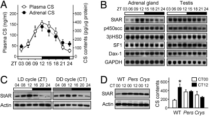
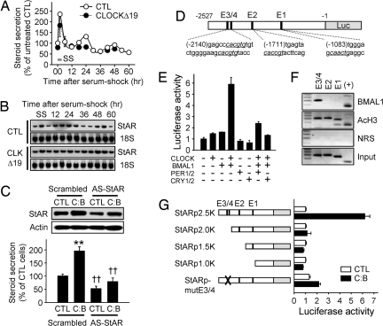
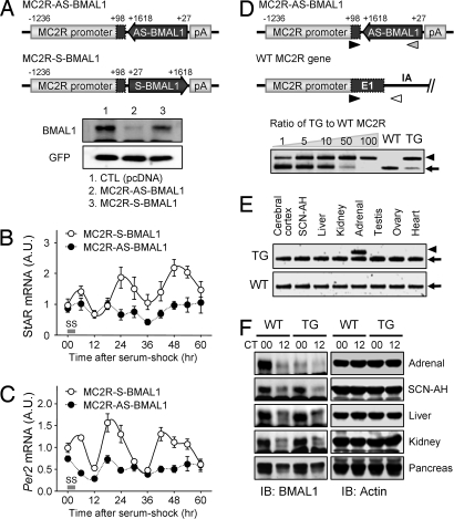
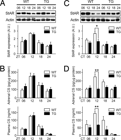
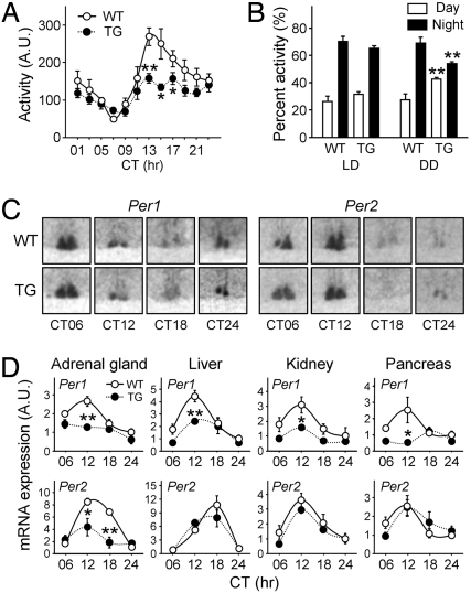
Glucocorticoid (GC) is an adrenal steroid with diverse physiological effects. It undergoes a robust daily oscillation, which has been thought to be driven by the master circadian clock in the suprachiasmatic nucleus of the hypothalamus via the hypothalamus-pituitary-adrenal axis. However, we show that the adrenal gland has its own clock and that the peripheral clockwork is tightly linked to steroidogenesis by the steroidogenic acute regulatory protein. Examination of mice with adrenal-specific knockdown of the canonical clock protein BMAL1 reveals that the adrenal clock machinery is required for circadian GC production. Furthermore, behavioral rhythmicity is drastically affected in these animals, together with altered expression of Period1, but not Period2, in several peripheral organs. We conclude that the adrenal peripheral clock plays an essential role in harmonizing the mammalian circadian timing system by generating a robust circadian GC rhythm.


Conflict of interest statement

The authors declare no conflict of interest.

Figures

Fig. 1.
Fig. 1.
Circadian rhythms of CS levels and adrenal StAR expression. (A) Daily profiles of CS in plasma (n = 8–9 per point) and adrenal lysates (n = 8–10) were expressed as mean ± SE. CS content in adrenal lysates was normalized with protein content. ZT, Zeitgeber time. (B) The mRNA profiles for the indicated steroidogenesis-related genes in adrenal gland and testis obtained by Northern blotting. StAR indicates steroidogenic acute regulatory protein; P450scc, P450 side-chain cleavage enzyme; 3βHSD, 3β-hydroxysteroid dehydrogenase; SF-1, steroidogenic factor-1; Dax-1, dosage-sensitive sex reversal adrenal hypoplasia congenital critical region on the X chromosome-1; and GAPDH, glyceraldehyde-3-phosphate dehydrogenase as an internal control. (C) Adrenal StAR protein levels under LD and DD conditions measured by immunoblotting. CT, Circadian time. (D) StAR protein level (Left) and adrenal CS content (Right) were examined in wild-type (WT), Per1−/−Per2−/− (Pers) and Cry1−/−Cry2−/− (Crys) mice (n = 6 for WT and Pers mice, and n = 3 for Crys mice; *, P < 0.05 between CT00 and CT12).
Fig. 2.
Fig. 2.
Transcriptional regulation of StAR expression by the cellular clock and the role of StAR in steroid production. (A) Adrenocortical Y1 cells were transfected with backbone plasmid (CTL) or the CMV promoter-driven CLOCKΔ19 plasmid (CLOCKΔ19). After serum shock (SS), hourly steroid secretion rates were measured and are expressed as percent (mean ± SE; n = 4–5). (B) With the same SS procedure, StAR mRNA levels were examined by Northern blotting. (C) Y1 cells were transfected with CTL or a 1:1 mixture of CLOCK and BMAL1-expressing constructs (C:B). Ten micrograms of scrambled (5′-GCTCTATGACTCCCAG-3′) or AS-StAR (5′-CATTTGGGTTCCACTC-3′) oligodeoxynucleotide (ODN) was included at the same time. Forty-eight hours after transfection, StAR protein levels (Upper) and steroid production (Lower) were measured (n = 4–5; **, P < 0.05 vs. CTL cells; ††, P < 0.05 vs. scrambled ODN-treated cells). (D) Schematic diagram of the mouse StAR promoter fused to the luciferase reporter (StARp2.5K). (E) Y1 cells were transfected with StARp2.5K in combination with various clock gene-expressing plasmids as indicated. Normalized luciferase activities are presented as mean ± SE of arbitrary unit (n = 6 for each group). (F) To reveal the binding of endogenous BMAL1 on the StAR promoter, chromatin immunoprecipitation assays were performed with anti-BMAL1 antibody, anti-acetylated histone H3 (AcH3) antibody, or preimmune normal rabbit serum (NRS). (+) indicates the functional E box-containing region on the D box-binding protein (DBP) promoter (48). (G) Effect of CLOCK:BMAL1 overexpression on luciferase activities driven by serially deleted or E box-mutated StAR promoters (n = 6–10 for each group).
Fig. 3.
Fig. 3.
Generation of MC2R-AS-BMAL1 TG mice. (A) Schematic diagrams of the MC2R-AS-BMAL1 and MC2R-S-BMAL1 constructs (Upper). pA indicates the poly(A) signal from the bovine growth hormone gene. Effect of pMC2R-AS-BMAL1 or pMC2R-S-BMAL1 on endogenous BMAL1 protein levels in Y1 cells (Lower). Cotransfected GFP was used as an internal control (CTL). (B and C) Y1 cells were transfected with MC2R-S-BMAL1 or MC2R-AS-BMAL1. After serum shock, StAR (B) and Per2 (C) mRNA levels were examined. Data were normalized with TATA-binding protein (TBP) and are expressed as mean ± SE arbitrary units (A.U.), where the mean value of the MC2R-S-BMAL1 group at 0 h was set at 1 (n = 3). (D) Schematic diagrams of the MC2R-AS-BMAL1 transgene and the endogenous MC2R gene (Upper). Primer binding sites for genotyping are indicated by arrowheads. The copy number of MC2R-AS-BMAL1 TG was determined by competitive PCR genotyping (Lower). The arrowhead indicates PCR products derived from the MC2R-AS-BMAL1, and arrows indicate those from the endogenous MC2R gene. (E) Adrenal gland-specific mRNA expression of AS-BMAL1 TG as revealed by competitive RT-PCR. Arrowhead indicates PCR products derived from MC2R-AS-BMAL1, and arrows from endogenous Bmal1 mRNA. (F) Adrenal gland-specific knockdown of BMAL1 expression. BMAL1 protein levels at circadian time (CT)00 and CT12 were examined in the indicated tissues by immunoblotting.
Fig. 4.
Fig. 4.
Dampened circadian CS rhythm in MC2R-AS-BMAL1 TG mice. WT and TG mice housed under 12 h of LD photoperiod (A and B) or DD conditions (C and D; 6–7 days after lights-off) were killed at ZT or CT 6, 12, 18, and 24 h. StAR protein levels in the adrenal glands were determined by immunoblotting (A and C; n = 4; *, P < 0.05 between WT and TG). (B and D) Adrenal CS contents (Upper) and plasma CS levels (Lower) in WT and TG mice were measured by RIA and are expressed as mean ± SE (n = 4–11 for DD and 4–6 for LD; **, P < 0.01 between WT and TG).
Fig. 5.
Fig. 5.
Behavioral rhythm and clock gene expression in MC2R-AS-BMAL1 TG mice. (A) Mean home-cage activities over the 7–14 days after the beginning of a DD cycle are presented as means ± SE, at 2-h intervals (n = 6 for each group; *, P < 0.05 and **, P < 0.01 between WT and TG). (B) Relative home-cage activities during rest (ZT or CT00–CT12) and activity periods (ZT or CT12–CT24) under LD or DD conditions. Activities under DD conditions were obtained during the second week of constant darkness. Data are presented as means ± SE % of total activity per day (n = 10 for each group; **, P < 0.01 vs. WT over the same period). (C and D) WT and TG mice housed under DD conditions for 6–7 days were killed at CT 6, 12, 18, and 24 h. (C) Per1 and Per2 expression in the SCN as revealed by in situ hybridization. (D) Per1 and Per2 mRNA expression profiles were obtained by real-time RT-PCR in the indicated organs. Data are normalized with TBP and expressed as means ± SE of A.U., where the mean WT value at CT24 is defined as 1 (n = 4–6, *, P < 0.05 and **, P < 0.01 vs. WT at the same time points).

References

    1. Wang M. The role of glucocorticoid action in the pathophysiology of the metabolic syndrome. Nutr Metab. 2005;2:3. - PMC - PubMed
    1. Buckingham JC. Glucocorticoids: Exemplars of multi-tasking. Br J Pharmacol. 2006;147:S258–S268. - PMC - PubMed
    1. Ko CH, Takahashi JS. Molecular components of the mammalian circadian clock. Hum Mol Genet. 2006;15:R271–R277. - PubMed
    1. Stratmann M, Schibler U. Properties, entrainment, and physiological functions of mammalian peripheral oscillators. J Biol Rhythms. 2006;21:494–506. - PubMed
    1. Balsalobre A, et al. Resetting of circadian time in peripheral tissues by glucocorticoid signaling. Science. 2000;289:2344–2347. - PubMed

Publication types

MeSH terms