. 2008 Dec 17;28(51):13742-53.
doi: 10.1523/JNEUROSCI.4844-08.2008.

Organization and postembryonic development of glial cells in the adult central brain of Drosophila

Affiliations

Organization and postembryonic development of glial cells in the adult central brain of Drosophila

Takeshi Awasaki et al. J Neurosci. .

Abstract

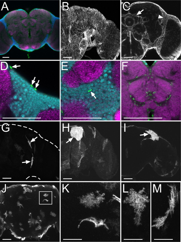
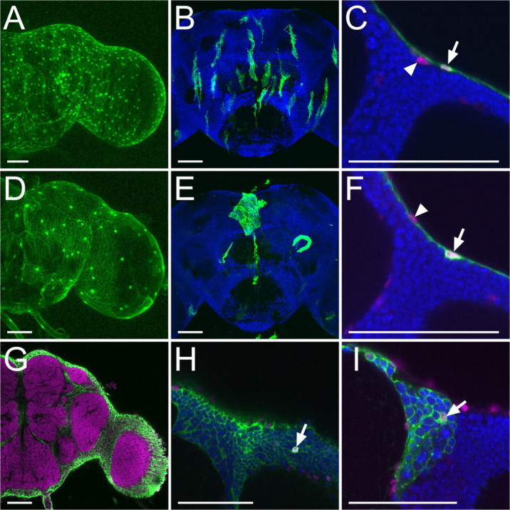
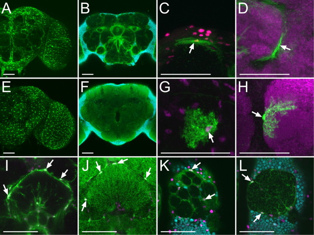
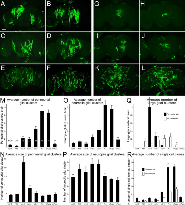
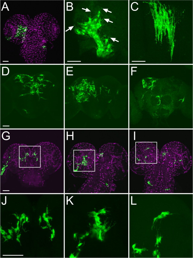
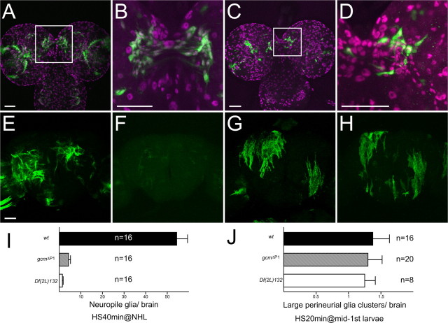
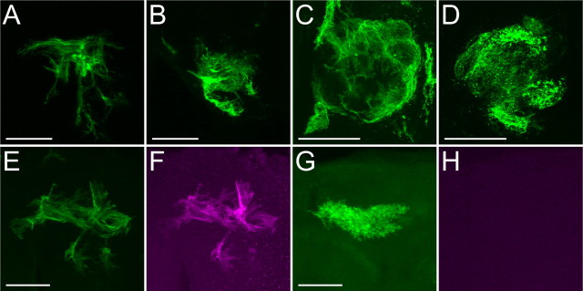
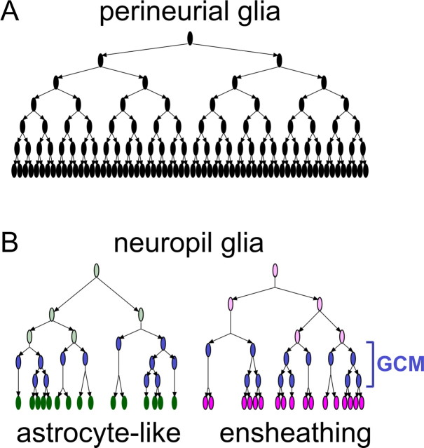
Glial cells exist throughout the nervous system, and play essential roles in various aspects of neural development and function. Distinct types of glia may govern diverse glial functions. To determine the roles of glia requires systematic characterization of glia diversity and development. In the adult Drosophila central brain, we identify five different types of glia based on its location, morphology, marker expression, and development. Perineurial and subperineurial glia reside in two separate single-cell layers on the brain surface, cortex glia form a glial mesh in the brain cortex where neuronal cell bodies reside, while ensheathing and astrocyte-like glia enwrap and infiltrate into neuropils, respectively. Clonal analysis reveals that distinct glial types derive from different precursors, and that most adult perineurial, ensheathing, and astrocyte-like glia are produced after embryogenesis. Notably, perineurial glial cells are made locally on the brain surface without the involvement of gcm (glial cell missing). In contrast, the widespread ensheathing and astrocyte-like glia derive from specific brain regions in a gcm-dependent manner. This study documents glia diversity in the adult fly brain and demonstrates involvement of different developmental programs in the derivation of distinct types of glia. It lays an essential foundation for studying glia development and function in the Drosophila brain.


Figures

Figure 1.
Figure 1.
Repo-positive glial cells in the adult central brain. A, Glial nuclei labeled by anti-Repo antibody (green), neuronal nuclei labeled by anti-Elav (blue), and neuropil labeled by nc82 (magenta). B, C, Glial membrane labeled with mCD8::GFP driven by repo-GAL4 on brain surface (B) and inner brain (C). Arrow and arrowhead show glial membrane forming borders among substructures of neuropil and brain cortex, respectively. D–F, Glial nuclei (green) on brain surface (D), cortex (E), and neuropil (F). Glial nuclei (arrows in D and E), neuronal nuclei, and neuropil were labeled as shown (A). G–I, Single cells of small surface glia (G), large surface glia (H), and cortex glia (I) labeled by FLP-out combined with repo-GAL4. J–M, Single cells of neuropil glia labeled by FLP-out combined with repo-GAL4. A dendritic and fibrous lamellar morphology of neuropil glia was labeled around neuropil (J). Two different types of neuropil glia arranged nearby (square in J) are shown at a high magnification (K). L, M, High-magnification images of a dendritic (L) and fibrous lamellar morphology (M) of neuropil glia. Scale bars: A–J, 50 μm; K–M, 25 μm.
Figure 2.
Figure 2.
Surface and cortex glia of adult brain. A–F, Perineurial glia (A–C, green) and subperineurial glia (D–F, green) labeled with UAS-GFP (A, C, D, F) and UAS-FRT-mCD8::GFP (FLP-out) (B, E), driven by NP6293 (A–C) and NP2276 (D–F), respectively. Glial nuclei and neuronal nuclei were labeled with anti-Repo (magenta) and anti-Elav (blue), respectively. Note that GAL4-negative, Repo-positive surface nuclei (arrowheads, C and F) were labeled under and over the layer of perineurial glia and subperineurial glia, respectively (arrows in C and F). G–I, Cortex glial cells (green) labeled with UAS-GFP driven by NP2222 (G, H) and with FLP-out combined with NP2222 (I). Neuropils were labeled with nc82 (magenta in G). Glial nuclei (magenta) and neuronal nuclei (blue) were labeled with anti-Repo and anti-Elav, respectively (H, I). Arrows show nuclei of cortex glia. Scale bars, 50 μm.
Figure 3.
Figure 3.
Two subtypes of neuropil glia of adult central brain. Ensheathing (A–D, I, K) and astrocyte-like (E–H, J, L) glia labeled with NP1243 and NP6520, respectively. Entire and single cells of neuropil glial subtype were labeled with UAS-GFP (A, E, and I–L), UAS-mCD8::GFP (B, F), and UAS-FLP-out (C, D, G, H), respectively. Arrows show nuclei of each subtype of glial cells labeled with anti-Repo (magenta in C, G, and I–L). Neuronal nuclei (blue) were labeled with anti-Elav (B, F, K, L) and neuropil (magenta) was labeled with nc82 (D, H). Scale bars, 50 μm.
Figure 4.
Figure 4.
Systematic MARCM analysis of glial proliferation during postembryonic development. A–L, Perineurial (A–F) and neuropil (G–L) glial cells labeled by heat-shock-induced mitotic recombination in mid-first-instar larvae (A, B), newly hatched larvae (G, H), early third-instar larvae (C, D, I, J) and early pupae (E, F, K, L). Scale bar, 50 μm. M–R, Quantification of glial cells that were labeled by MARCM system with heat-shock-induced mitotic recombination at different developmental stages (x-axis). Average number (M, O) and size (N, P) of perineurial glia (M, N) and neuropil glia (O, P) clusters that were induced at different developmental stage (y-axis). Q, Average number of large perineurial glia (>10 cells) and neuropil glia (>8) clusters per brain. R, Average number of single cell clones of perineurial and neuropil glia. Same samples were used for analysis in MR. Numbers of examined brains are shown in M. Genotypes: hs-FLP/repo-GAL4; FRTG13, UAS-mCD8::GFP/FRTG13, tubP-GAL80, repo-GAL80 (A–L) and hs-FLP/repo-GAL4; FRTG13, UAS-nlsGFP/FRTG13, tubP-GAL80, repo-GAL80 (M–R).
Figure 5.
Figure 5.
Precursors of perineurial and neuropil glia. Perineurial glia cluster in a wandering larva (A, B) and adult (C), which were labeled by MARCM system with heat-shock-induced mitotic recombination in mid-first-instar larvae. Arrows show filopodial processes extending from larval perineurial glia. Multiple neuropil glia clusters in adults (D–F) and wandering larvae (G–L), which were labeled by MARCM with long heat-shock (40 min) induced mitotic recombination in NHL. Note that labeled cells were localized in posterior-medial (G, J), lateral (H, K), and dorsal (I, L) interface between brain cortex and neuropil. J–L, High-magnification images of G–I, respectively. Glial nuclei were labeled with anti-Repo (magenta, A and G–I). Scale bars, 50 μm.
Figure 6.
Figure 6.
Function of gcm on postembryonic development of perineurial and neuropil glia. A, B, Cells labeled with GFP driven by gcm-GAL4 in a wandering larva. C, D, Cells labeled by MARCM system with gcm-GAL4. Mitotic recombination was induced in NHL. Glial nuclei were labeled with anti-Repo antibody (magenta, A–D). High-magnification images of A and C are shown in B and D, respectively. Note that gcm-positive cells were labeled with anti-Repo antibody (B, D). E–H, Wild-type (E, G) and gcm ΔP1 (F, H) MARCM clones of neuropil glia (E, F) induced at NHL and perineurial glia (G, H) induced at mid-first-instar larvae, respectively. I, J, Quantification of neuropil glia (I) and perineurial glia (J) that were labeled by MARCM system. Average number of neuropil glia per brain (I) and average number of large perineurial glia cluster (>10) per brain were examined in wt, gcm ΔP1, and Df(2L)132 clones. Scale bars, 50 μm. Genotypes: gcm-GAL4/UAS-GFP (A, B); hs-FLP/+; gcm-GAL4, FRTG13, UAS-nlsGFP/FRTG13, tubP-GAL80, repo-GAL80 (C, D); hs-FLP, UAS-mCD8::GFP/repo-GAL4; FRT40A, tubP-GAL80/FRT40A or FRT40A, gcm ΔP1 (E–H); and hs-FLP/repo-GAL4; FRT40A, tubP-GAL80/FRT40A or FRT40A, gcm ΔP1 or FRT40A, Df(2L)132; UAS-nlsGFP/+ (I, J).
Figure 7.
Figure 7.
Neuropil glial clones. Ensheathing glial clones (A, C, E, F) and astrocyte-like glial clones (B, D, G) labeled by standard MARCM (A–D) and dual-expression control MARCM (E–H). Note that C and D show glial clones located in the antennal lobe. Whereas the ensheathing glial clone was labeled by both rCD2::GFP driven by repo-LexA::GAD (green, E) and mCD8::RFP driven by NP6520 (magenta, F), the astrocyte-like glial cone was labeled by only rCD2::GFP driven by repo-LexA::GAD (green, G), but not by mCD8::RFP driven by NP6520 (magenta, H). Scale bars, 50 μm. Genotypes: hs-FLP/repo-GAL4; FRTG13, UAS-mCD8::GFP/FRTG13, tubP-GAL80, repo-GAL80 (A–D) and hs-FLP/repo-LexA::GAD; FRTG13/FRTG13, tubP-GAL80, repo-GAL80; NP6520/UAS-mCD8::RFP, LexA-operator-rCD2::RFP (E–H).
Figure 8.
Figure 8.
Schematic model of postembryonic glial proliferation. A, B, Perineurial glia (A) and two subtypes of neuropil glia (B).

Similar articles

Cited by

References

    1. Awad TA, Truman JW. Postembryonic development of the midline glia in the CNS of Drosophila: proliferation, programmed cell death, and endocrine regulation. Dev Biol. 1997;187:283–297. - PubMed
    1. Awasaki T, Ito K. Engulfing action of glial cells is required for programmed axon pruning during Drosophila metamorphosis. Curr Biol. 2004;14:668–677. - PubMed
    1. Awasaki T, Tatsumi R, Takahashi K, Arai K, Nakanishi Y, Ueda R, Ito K. Essential role of the apoptotic cell engulfment genes draper and ced-6 in programmed axon pruning during Drosophila metamorphosis. Neuron. 2006;50:855–867. - PubMed
    1. Bainton RJ, Tsai LT, Schwabe T, DeSalvo M, Gaul U, Heberlein U. moody encodes two GPCRs that regulate cocaine behaviors and blood-brain barrier permeability in Drosophila . Cell. 2005;123:145–156. - PubMed
    1. Beckervordersandforth RM, Rickert C, Altenhein B, Technau GM. Subtypes of glial cells in the Drosophila embryonic ventral nerve cord as related to lineage and gene expression. Mech Dev. 2008;125:542–557. - PubMed

Publication types

MeSH terms

LinkOut - more resources