. 2009 Apr;80(4):696-706.
doi: 10.1095/biolreprod.108.071589. Epub 2008 Dec 17.

The novel epididymis-specific beta-galactosidase-like gene Glb1l4 is essential in epididymal development and sperm maturation in rats

Affiliations

The novel epididymis-specific beta-galactosidase-like gene Glb1l4 is essential in epididymal development and sperm maturation in rats

Wei Zhen et al. Biol Reprod. 2009 Apr.

Abstract

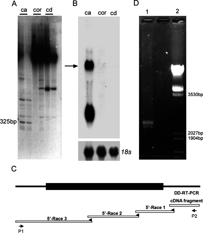
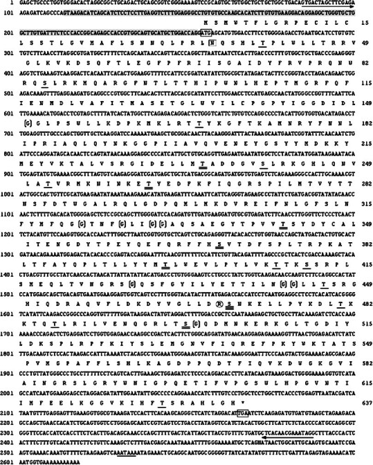
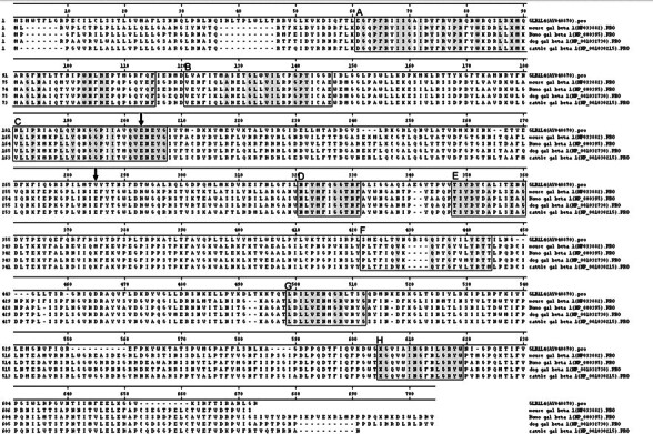
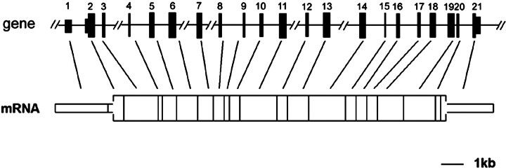
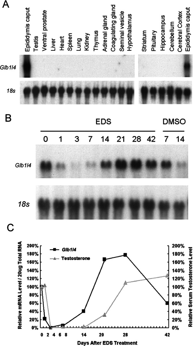
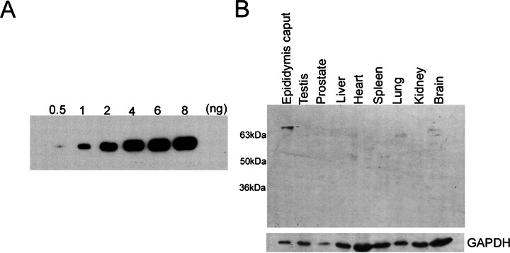
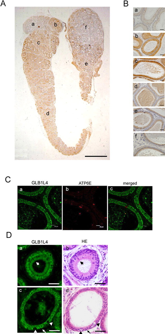
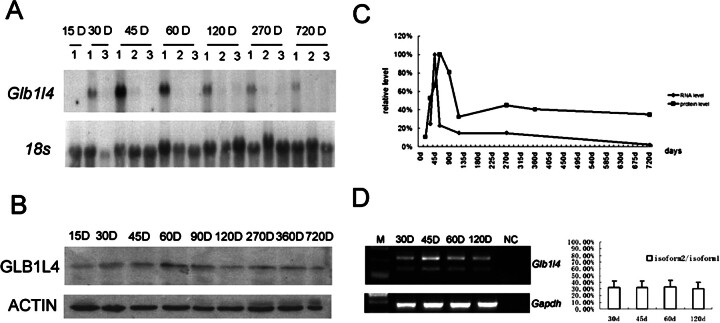
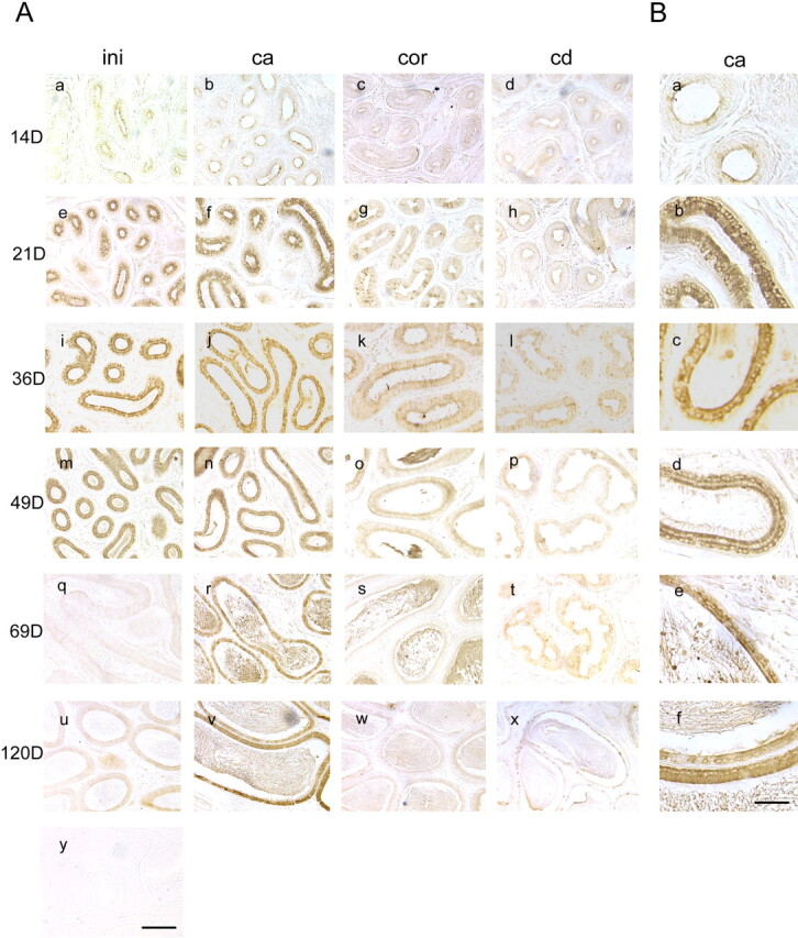
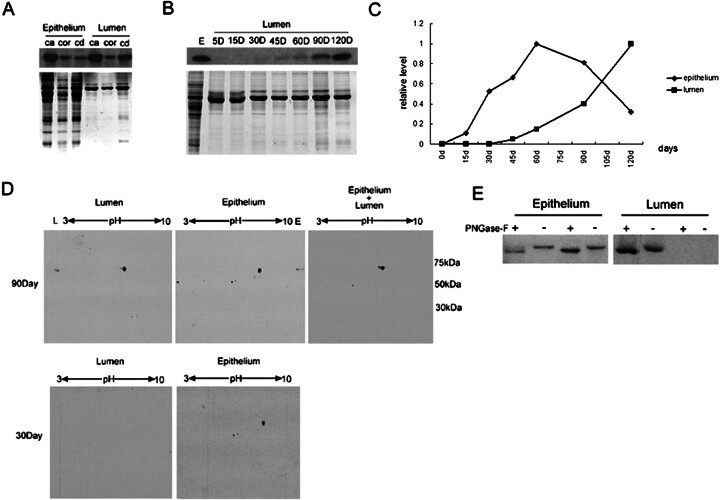
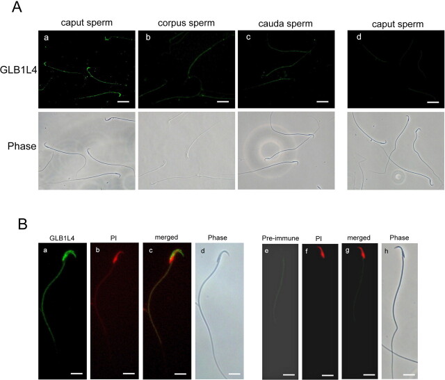
We describe a novel epididymis-specific cDNA named Glb1l4, which was isolated from rat epididymis by differential display of mRNAs. Glb1l4 cDNA contains 2607 nucleotides and encodes a 637-amino acid protein with 50% similarity to mouse beta-galactosidase. The gene is located on chromosome 8q13, spanning 21 exons. Northern blot analysis reveals that Glb1l4 is specifically expressed in the caput region of epididymis and upregulated by androgen. A specific polyclonal antiserum against the N-terminal peptide of GLB1L4 has been produced. Western blot analysis and immunohistochemistry assay reveal that GLB1L4 is specifically expressed in the principal cells of the caput epididymis. Interestingly, its expression peaks at Postnatal Day 45 in mRNA level and at Postnatal Day 60 in protein level while the epididymis column cells undergo differentiation. Moreover, within this very period this secretory protein is confined inside the cell with a change of subcellular distribution pattern, which implies its important roles in the cell differentiation process. Only after the epididymal epithelium differentiation is completed and the spermatozoa enter the epididymal lumen is the GLB1L4 secreted into the luminal fluid and bound on the sperm head. Our results suggest that GLB1L4 may play various roles in principal cell differentiation and sperm maturation.


Figures

Fig. 1
Fig. 1
Cloning of the full length of rat Glb1l4. A) A DD-RT-PCR analysis of differentially expressed mRNAs in the epididymides of rats. Shown are differentially expressed transcripts obtained with combinations of upper primer 503 (CTTTCTACCC) and lower primer (T11CA) in three regions of the rat epididymis: caput (ca), corpus (cor), and cauda (cd). A 325-bp fragment is principally expressed in the caput region. B) The fragment obtained from A was used as a probe in three regions of the rat epididymis to identify mRNAs hybridized by Northern blot analysis. The Glb1l4 mRNA is indicated by an arrow. C) Diagram of cloning for Glb1l4 cDNA by 5′-RACE. Based on the 325-bp sequence of the DD-RT-PCR fragment, gene-specific primers were designed and the sequence toward the 5′ end was extended with 456, 593, and 1232, respectively, through three consecutive 5′-RACE reactions. D) Amplification of full-length Glb1l4 cDNA by RT-PCR. Two bands were amplified by the specific primers P1 and P2. The products of RT-PCR were loaded in lane 1, and DNA size markers were loaded in lane 2.
Fig. 2
Fig. 2
The cDNA and deduced amino acid sequences of Glb1l4. The initial and terminal codons are boxed. The polyadenylation signal (AATAAA) is underlined. The sequences for primers P1 and P2 are underlined with arrows. The 146-bp sequence deleted in the short form is shadowed gray. The N-glycosylation (N35) site is boxed; 11 protein kinase C phosphorylation sites are underlined; six casein kinase II phosphorylation sites are double underlined; S470, which may be both a protein C kinase phosphorylation site and a casein kinase II phosphotylation site, is triple underlined; nine N-myristoylation sites are bracketed; and one tyrosine kinase phosphorylation site is circled. Numbers on the left and on the right correspond to nucleotide and amino acid positions, respectively.
Fig. 3
Fig. 3
Amino acid sequence alignment of rat GLB1L4 and members of the beta-galactosidase family from mice, humans, dogs, and cattle. The boxes show the conserved regions of beta-galactosidase (Glyco_hydro_35 block number IPB001944A–IPB001944H). The amino acid sequences in eight conserved regions of five proteins are shadowed in gray. The active sites of catalytic proton donor E205 and catalytic nucleophile E279 are indicated by arrows. The catalytic nucleophile site E279 is replaced by V279 in rat GLB1L4 protein.
Fig. 4
Fig. 4
The genomic structure of the rat Glb1l4 gene. The Glb1l4 gene diagram shows the relative lengths of exons 1–21 and intervening introns. Line breaks indicate that the introns have lengths greater than 2 kb and are not drawn to scale. The open box represents the ORF of Glb1l4 mRNA. The narrow bar represents the 5′ and 3′ untranslated regions of the Glb1l4 mRNA.
Fig. 5
Fig. 5
Tissue distribution and androgen manipulation of rat Glb1l4 mRNA analysis by Northern blot analysis. A) Total RNAs (20 μg/lane) from various tissues were used for Northern blot analysis. A specific band was detected in the caput epididymis only. The 18s rRNA was used as internal control. B) Testosterone increases Glb1l4 expression in the epididymis. Northern blot analyses were performed with total rat epididymis RNA (20 μg/lane) from pooled tissues (n = 5 for each time point, expressed in days) following EDS or DMSO (vehicle control) treatment and were hybridized with a Glb1l4 probe. The blot was reprobed for 18s ribosomal RNA to verify RNA integrity and equal loading. C) Quantitative analysis of Glb1l4 mRNA and serum testosterone following EDS treatment confirmed that Glb1l4 mRNA levels were correlated with changes in serum testosterone. Results are means from two independent experiments. Values were normalized to levels in control animals prior to EDS treatment (Day 0) and then were plotted as relative fold increases.
Fig. 6
Fig. 6
The sensitivity and specificity of the two polyclonal antisera against the N-terminal peptide of GLB1L4 were analyzed by Western blot analysis. In A, 0.5, 1, 2, 4, 6, and 8 ng of N-terminal antigen peptide were loaded. The antiserum can detect as low as 0.5 ng of antigen by Western blot analysis. B) Protein extracts (40 μg/lane) from various tissues were analyzed by Western blot analysis. GAPDH was used as an internal control.
Fig. 7
Fig. 7
The region-specific and cell-specific localization of rat GLB1L4 in the rat epididymis. A) The regional expression pattern of GLB1L4 in the whole epididymis of a 120-day-old rat. Bar = 2 mm. B) Magnified photographs for individual fields of A: the initial segment (a), the proximal caput (b), the caput (c), the corpus (d), the distal corpus (e), and the cauda (f). Bars = 50 μm. C) Immunofluorescence detection of GLB1L4 (a, FITC labeled), ATP6E (b, rhodamine labeled for clear cells), and their colocalization (c). Bars = 20 μm. D) Immunofluorescence detection of GLB1L4 (a and c) and HE staining (b and d) of the same section, showing that the narrow cell (large arrows), halo cell (arrowheads), and basal cell (small arrows) are not reactive. Bars = 50 μm.
Fig. 8
Fig. 8
Expression of Glb1l4 in rat epididymis during the entire life span in days (D). A) Northern blot analysis for Glb1l4 mRNA and 18s rRNA from the caput region (1), the corpus region (2), and the cauda region (3) during development. B) Western blot analysis of GLB1L4 and ACTIN proteins during development. D, days. C) Quantitative analysis of the relative mRNA and protein expression levels in rat epididymides at different ages. RNA and protein were pooled from three animals per group. D) Left: RT-PCR of two isoforms of Glb1l4 mRNA expression in rat caput epididymis from 30, 45, 60, and 120 days (D) after birth. Gapdh was used as internal control. NC, negative control. Right: semiquantitative analysis of the ratio of isoform2:isoform1 in rat epididymis at different ages. d, days.
Fig. 9
Fig. 9
The spatial and temporal expressions of rat GLB1L4 in rat epididymis. The GLB1L4 protein localization in different regions of the rat epididymis at different ages was determined by immunohistochemistry. The ages in days (D) are shown on the left, and the regions of the epididymis are shown at the top. Panel y, as detected by preimmune serum, served as negative control. ini, the initial segment; ca, the caput region; cor, the corpus region; cd, the cauda region. Bar = 100 μm. B) Each panel is a highly magnified image of the caput epididymis as shown in A (i.e., Ab, Af, Aj, An, Ar, and Av). Bar = 25 μm.
Fig. 10
Fig. 10
Comparison of GLB1L4 in the rat epididymal epithelium and lumen by Western blot analysis. A) Western blot analysis of GLB1L4 from rat epididymal epithelium and lumen protein extracts of the caput (ca), corpus (cor), and cauda (cd) regions. Protein (30 μg) was loaded in each lane. The same volume of protein was separated by electrophoresis and stained by Coomassie blue to show the equal loading. B) Western blot analysis of GLB1L4 from epididymal lumen protein during development. Protein (20 μg) was loaded in each lane. The same volume of protein was separated by electrophoresis and stained by Coomassie blue to show the equal loading. E, epididymis; D, days. C) Quantitative analysis of the relative protein expression levels in epididymal epithelium and lumen fluid at different ages in days (d). Protein extracts were pooled from three animals per group. D) Lumen (100 μg), epididymal epithelium (100 μg), and epithelium plus lumen (200 μg epithelium and 100 μg lumen) proteins were separated by 2D gel electrophoresis and detected using anti-GLB1L4 antisera. Molecular mass is indicated on the right (kDa), and pI values are shown on the top. E) The change in molecular mass of mature (90-day-old; left) and adolescent (30-day-old; right) rat GLB1L4 before (−) and after (+) deglycosylation by PNGase-F. Epithelium, epididymal epithelium protein; Lumen, lumen fluid protein.
Fig. 11
Fig. 11
Rat GLB1L4 localization on spermatozoa derived from the caput, corpus, and cauda epididymal regions by immunofluorescence. A) Immunolocalization of rat GLB1L4 (FITC labeled) on spermatozoa isolated from different epididymal regions: (a) caput spermatozoa, (b) corpus spermatozoa, (c) cauda spermatozoa, and (d) caput spermatozoa as detected by preimmune serum as negative control. Bottom row of photographs are phase-contrast images of the spermatozoa shown in the photographs in the upper row. Bars = 10 μm. B) Highly magnified images of the GLB1L4 protein localization on caput spermatozoa: (a) the immunofluorescence of rat GLB1L4 (FITC labeled); (b) spermatozoa nuclear detected by propidium iodide (PI; rhodanmine labeled); (c) the merged photographs of a and b; (d) phase-contrast view of sperm in a. e) Caput spermatozoa detected by preimmune serum as negative control; (f) spermatozoa nuclear detected by PI; (g) the merged photographs of e and f; and (h) phase-contrast view of sperm in e. Bars = 5 μm.

References

    1. Rodriguez CM, Kirby JL, Hinton BT. The development of the epididymis.In:Robaire B, Hinton BT. The Epididymis: From Molecules to Clinical Practice. New York: Kluwer Academic/Plenum Publishers; 2002:251–267.
    1. Connwall GA, Hann SR. Specialized gene expression in the epididymis. J Androl 1995; 16:379–383. - PubMed
    1. Hamilton DW. Structure and function of the epithelium lining the ductuli efferents, ducts epididymis and ductus deferens in the rat.In:Greep RO, Astwood EB. Handbook of Physiology, vol. 5. Baltimore: Waverly Press; 1975:259–383.
    1. Miler RJ, Killian GJ. Morphometric analysis of the epididymis from normal and vasectomized rats. J Androl 1987; 8:279–291. - PubMed
    1. Wong PYD, Au CL, Ngai HK. Electrolyte and water transport in rat epididymis: its possible role in sperm maturation. Int J Androl 1978; 1:600–628.

Publication types

MeSH terms