. 2009 Feb;20(2):333-43.
doi: 10.1681/ASN.2008070795. Epub 2008 Dec 17.

Recruitment of podocytes from glomerular parietal epithelial cells

Affiliations

Recruitment of podocytes from glomerular parietal epithelial cells

Daniel Appel et al. J Am Soc Nephrol. 2009 Feb.

Abstract

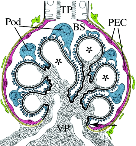
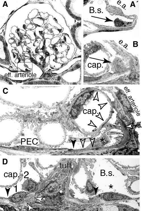
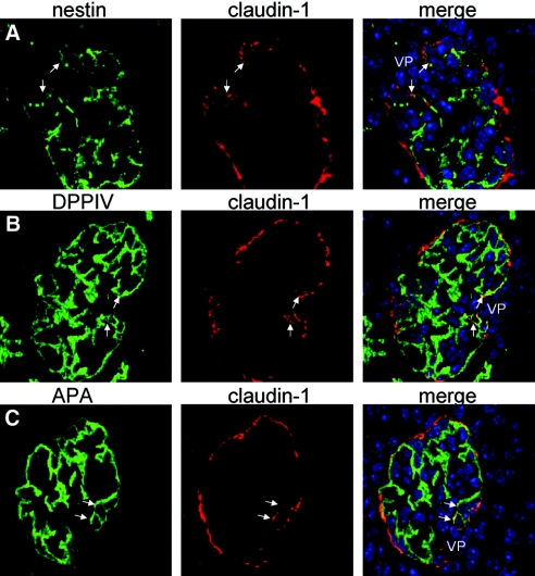
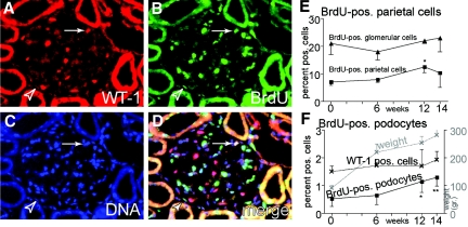
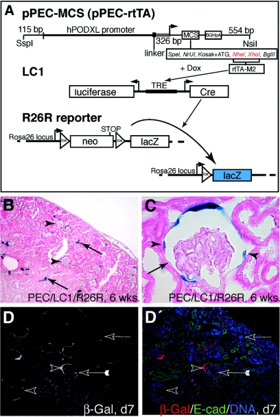
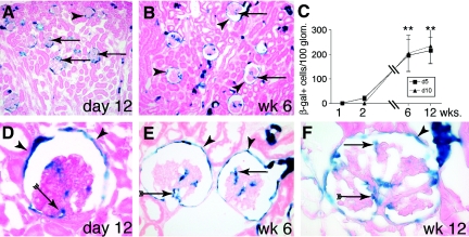
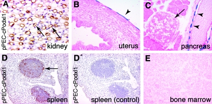
Loss of a critical number of podocytes from the glomerular tuft leads to glomerulosclerosis. Even in health, some podocytes are lost into the urine. Because podocytes themselves cannot regenerate, we postulated that glomerular parietal epithelial cells (PECs), which proliferate throughout life and adjoin podocytes, may migrate to the glomerular tuft and differentiate into podocytes. Here, we describe transitional cells at the glomerular vascular stalk that exhibit features of both PECs and podocytes. Metabolic labeling in juvenile rats suggested that PECs migrate to become podocytes. To prove this, we generated triple-transgenic mice that allowed specific and irreversible labeling of PECs upon administration of doxycycline. PECs were followed in juvenile mice beginning from either postnatal day 5 or after nephrogenesis had ceased at postnatal day 10. In both cases, the number of genetically labeled cells increased over time. All genetically labeled cells coexpressed podocyte marker proteins. In conclusion, we demonstrate for the first time recruitment of podocytes from PECs in juvenile mice. Unraveling the mechanisms of PEC recruitment onto the glomerular tuft may lead to novel therapeutic approaches to renal injury.


Figures

Figure 1.
Figure 1.
Renal glomerulus. The glomerular epithelium consists of PECs (red) and podocytes (Pod; blue), which reside on the capillary convolute. Both epithelia adjoin directly at the vascular pole (VP; arrow). At the tubular pole (TP), the parietal epithelium is connected to the epithelium of the proximal tubule. In male mice, this transition from PECs to proximal tubular cells often occurs within the glomerulus. The glomerular basement membrane (black) forms a continuous barrier between the glomerular epithelium and the endocapillary compartment that contains mesangial cells (shaded) and endothelial cells of the glomerular capillaries (*). Primary urine is filtered across the three-layered filtration barrier (endothelial cells, glomerular basement membrane, and Pod) into Bowman's space (BS).
Figure 2.
Figure 2.
Transitional cells at the rat parietal cell–Pod interface. (A) At the VP, epithelial cells with features of parietal cells and Pod can be regularly observed. (A′) Early transitional stage (characteristics: glomerular basement membrane, cytoplasmic vesicles; higher magnification of box in A); e.a., efferent arteriole. (B) Late transitional stage (characteristics: upright cell body, large lobulated nucleus); cap., capillary lumen. (C) Partial formation of foot processes within a “late” transitional cell (*) at the vascular stalk. The intercellular junction between the last parietal cell (PEC) and the transitional cell is marked by a filled arrowhead. The transition from a parietal cell basement membrane to a glomerular basement membrane also occurs at this site. The transitional cell projects extensions onto the base of Bowman's capsule as well as onto a capillary (cap.) without forming foot processes (open arrowheads). A third projection extends onto the vascular stalk and forms typical foot processes with a slit diaphragm (arrow). (D) Grazing section along the vascular stalk. Sequential stages of transitional cells at the parietal/Pod interface (1, early stage; 2, later stage). The intercellular junction toward the last PEC (*) is marked by arrowheads, the transition from a parietal to a glomerular basement membrane is marked by open arrows (A through C, transmission electron micrographs of adult Sprague-Dawley rats).
Figure 3.
Figure 3.
Transitional cells coexpress PEC and Pod marker proteins. (A through C) Normal mouse frozen kidney sections from 10-d-old mice were co-stained with an antiserum specific for claudin-1 (PECs, red) and the Pod markers (green) nestin (A), dipeptidyl peptidase 4 (DPPIV; B), and aminopeptidase A (APA; C). At the VP, claudin-1–positive cells could regularly be observed. These cells at the vascular stalk coexpressed Pod marker proteins (arrows, nuclei are stained blue; immunofluorescent labelings of 2-μm sections analyzed by confocal microscopy).
Figure 4.
Figure 4.
Metabolic labeling of rat PECs. After weaning, 75-g female Sprague-Dawley rats were labeled with BrdU over 14 d and followed up to 14 wk. (A through D) Representative triple-immunofluorescent staining for the nuclear Pod marker protein WT-1 (red; A), BrdU (green; B), and DNA (blue; C) 12 wk after BrdU labeling. (D) Merged image (phase contrast image not shown). Arrowhead, BrdU-labeled PEC along the inner aspect of Bowman's capsule; arrow, BrdU-labeled WT-1–positive Pod nucleus on the glomerular tuft. (E) BrdU labeling persisted in glomerular cells (mainly mesangial and endothelial cells) without a significant increase over time (▴). Shortly after weaning, 7% of all PECs were metabolically labeled with BrdU. Over time, BrdU labeling of PECs increased significantly, most likely as a consequence of ongoing proliferation and self-regeneration of this cell population (*P = 0.02, one-sided ANOVA test). (F) BrdU-positive Pod were detected significantly more often at 12 or 14 wk after metabolic labeling (*P = 0.03, **P < 0.01, one-sided ANOVA test; n = 3 animals per time point, 300 glomeruli per animal). During the observation period, adolescent rats more than quadrupled their body weights (gray line). No significant increase in Pod numbers (WT-1–positive cells) relative to all glomerular cells was observed.
Figure 5.
Figure 5.
Identification of a PEC-specific promoter. (A) Map of the pPEC-cPodxl1 transgene. A 3-kb hybrid of the human and rabbit podocalyxin (hPODXL1) promoter (parietal cell promoter, pPEC) was used to drive expression of rabbit podocalyxin (cPodxl1) in transgenic mice. BGHpA, bovine growth hormone polyadenylation signal. (B through D) Transgene expression within the renal cortex. (B) In adult pPEC-cPodxl1–transgenic mice, rabbit podocalyxin was expressed exclusively in PECs of the renal cortex (arrowheads). (C) Transgene expression was restricted to PECs (arrowheads) and did not extend into the S1 segment of proximal tubular cells, which in male mice extends into Bowman's capsule (arrows). No labeling of Pod was observed (immunohistologic staining using anti-rabbit podocalyxin mouse mAb 4B3). (D and E) In newborn pPEC-cPodxl1 mice, the parietal promoter was active exclusively in mature PECs of the capillary loop stage or mature glomeruli (arrowheads). No transgene expression was observed in earlier developmental stages (e.g., S-shaped bodies [arrow]).
Figure 6.
Figure 6.
Genetic tagging of PECs in a triple-transgenic doxycycline-inducible rtTA mouse line. (A) pPEC-MCS, the rabbit podocalyxin cDNA, was replaced by a multiple cloning site (MCS; available restriction sites indicated). The enhanced tetracycline reverse transactivator (rtTA-M2) was cloned into Nhe1/Xho1 (red). pPEC-rtTA–transgenic mice were generated by pronuclear injection and mated to LC1 mice expressing Cre recombinase and luciferase under the control of tetracycline-responsive elements (TRE), which can be activated by rtTA-M2 in the presence of doxycycline and R26R: This reporter line irreversibly expresses β-gal (LacZ) under the control of the ubiquitous ROSA26 locus only after Cre excision of an interposed floxed neomycin cassette (neo; acting as a stop signal) has occurred. (B) Genetic labeling of PECs using doxycycline in 6-wk-old triple-transgenic pPEC-rtTA mice (pPEC-rtTA/LC1/R26R; 9 wk of age). Sporadic labeling was observed in some tubular cells of the renal cortex (arrows). (C) After induction, Cre recombination occurred in 72% of all PECs (arrowheads, labeled PEC; arrow, unlabeled PEC). (D and D′) Cre recombination was induced in triple-transgenic mice 5 d after birth (d5), when nephrogenesis still persists. Two days after doxycycline administration (d7), specific genetic labeling of PECs was verified by immunofluorescent staining (arrowheads). In 1 to 2% of all glomeruli, labeled cells on the glomerular tuft were observed (arrow with tails, confocal triple-immunofluorescent labeling; red, β-gal [genetic marker for PECs]; green, E-cadherin (proximal tubular cell marker); blue, DNA).
Figure 7.
Figure 7.
Recruitment of PECs onto the glomerular tuft in adolescent pPEC-rtTA/LC1/R26R mice. (A) After genetic tagging of PECs 5 d after birth, β-gal–positive cells (arrows) can be detected on glomerular tufts on day 12. (B) Six weeks after birth, genetically tagged cells are present within most glomeruli (arrows) of the outer cortex as well as close to the medulla. Genetic labeling persists in PECs (arrowheads). (C) Statistical analysis of β-gal–positive cells per 100 glomeruli over time in triple-transgenic PEC-TETon mice induced 5 (d5) or 10 d after birth (d10). A similar increase of β-gal–positive cells over time was observed in both groups (**P < 0.01 ANOVA; n = 5 for each time point). (D through F) Genetic labeling persists in PECs (arrowheads) 12 d (D) and 6 and 12 wk (E and F) after doxycycline administration. β-Gal–positive cells were identified close to the VP (arrow with tails) as well as projecting into the periphery of the glomerulus (arrow). (F) Occasionally, glomeruli with up to 20 β-gal–positive cells were observed at 12 wk of age (arrowheads, labeled PECs; A, B, and D through F, X-gal/eosin staining on 6-μm cryosections).
Figure 8.
Figure 8.
β-Gal–positive cells on the glomerular tuft are fully differentiated Pod. (A) Double-immunofluorescent staining for β-gal (red) and the Pod marker protein nephrin (green) in 6-wk-old triple-transgenic PEC-TETon mice induced with doxycycline at the age of 5 d. PECs expressed constitutively β-gal (open arrowheads). β-Gal–positive cells on the vascular tuft were exclusively Pod as demonstrated by nephrin coexpression (green, arrows). Transitional cells, located close to the vascular stalk, were genetically labeled (β-gal positive) and expressed low to intermediate levels of the Pod marker protein nephrin (filled arrowheads). (B) No co-localization of β-gal–positive cells (red, arrow) with the endothelial cell marker vWF (green, arrowheads) was observed in the mice described. Interstitial capillaries are marked in B (vWF) (open arrowhead). (C) β-Gal–positive Pod derived from PECs coexpress WT-1 (arrow, open arrowheads, β-gal positive/WT-1 negative PEC nuclei). Panel shows an enzymatic β-gal staining (in blue)/immunohistochemical 3-amino-9-ethyl-carbazole (AEC) anti–WT-1 (in red) double stainings.
Figure 9.
Figure 9.
Activity of the parietal cell promoter. (A) Within the kidney, activity of the PEC promoter was also observed within the thin limb of the loop of Henle (arrows, pPEC-cPodxl1 transgenic mouse; brown, anti-rabbit podocalyxin staining, hematoxylin counter staining). (B) Mesothelial cells lining the peritoneal cavity on the uterus were genetically labeled in a mosaic manner (arrowhead). (C) Cre recombination occurred within the epithelium of the pancreatic ducts (arrowheads) but not within glandular cells or pancreatic islets (arrow). (D and D′) Activity of the parietal cell promoter within follicles of the spleen (arrow) was visualized by immunohistology in pPEC-cPodxl1–transgenic mice (D, anti-rabbit podocalyxin in brown; D′ control using irrelevant primary antibody). (E) No evidence for Cre recombination was observed within cells of the bone marrow of mice more than 8 mo after induction (B, C, and E, pPEC-rtTA/LC1/R26R mouse, X-gal/eosin staining on cryosections).

Comment in

References

    1. US Renal Data System: USRDS 2007 Annual Report, Bethesda, National Institutes of Health, National Institute of Diabetes and Digestive and Kidney Diseases, 2007
    1. Wharram BL, Goyal M, Wiggins JE, Sanden SK, Hussain S, Filipiak WE, Saunders TL, Dysko RC, Kohno K, Holzman LB, Wiggins RC: Podocyte depletion causes glomerulosclerosis: Diphtheria toxin-induced podocyte depletion in rats expressing human diphtheria toxin receptor transgene. J Am Soc Nephrol 16: 2941–2952, 2005 - PubMed
    1. Novick AC, Gephardt G, Guz B, Steinmuller D, Tubbs RR: Long-term follow-up after partial removal of a solitary kidney. N Engl J Med 325: 1058–1062, 1991 - PubMed
    1. Kriz W, Gretz N, Lemley KV: Progression of glomerular diseases: Is the podocyte the culprit? Kidney Int 54: 687–697, 1998 - PubMed
    1. Hara M, Yamamoto T, Yanagihara T, Takada T, Itoh M, Adachi Y, Yoshizumi A, Kawasaki K, Kihara I: Urinary excretion of podocalyxin indicates glomerular epithelial cell injuries in glomerulonephritis. Nephron 69: 397–403, 1995 - PubMed

Publication types