. 2009 Mar;75(5):526-35.
doi: 10.1038/ki.2008.602. Epub 2008 Dec 17.

Renal ischemia-reperfusion leads to long term infiltration of activated and effector-memory T lymphocytes

Affiliations

Renal ischemia-reperfusion leads to long term infiltration of activated and effector-memory T lymphocytes

Miguel Ascon et al. Kidney Int. 2009 Mar.

Abstract

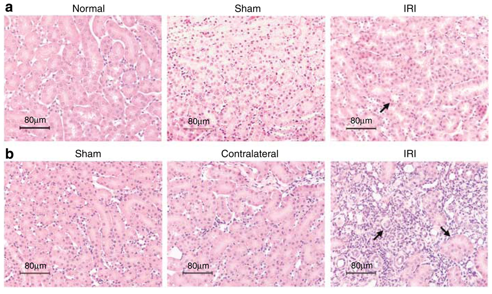
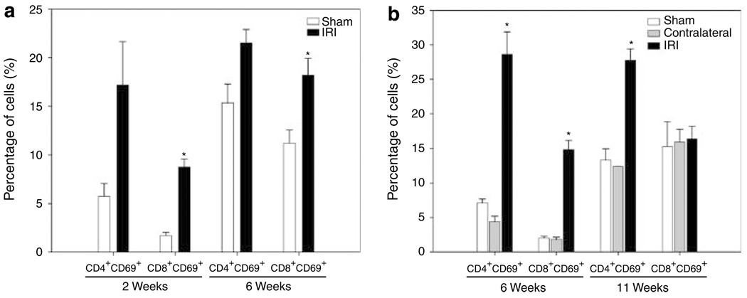
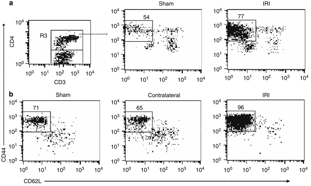
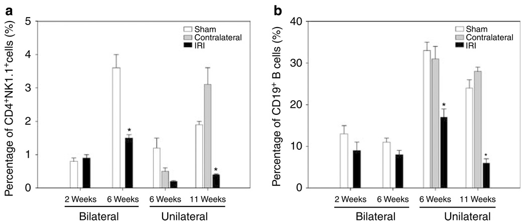
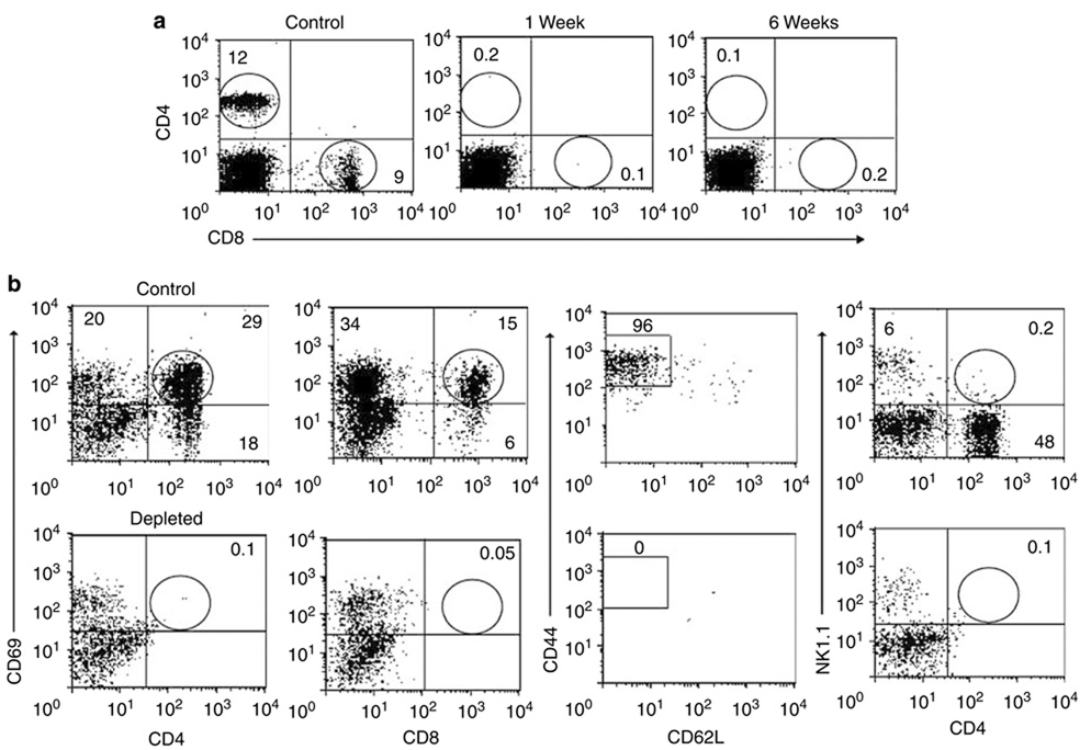
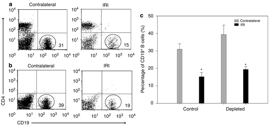
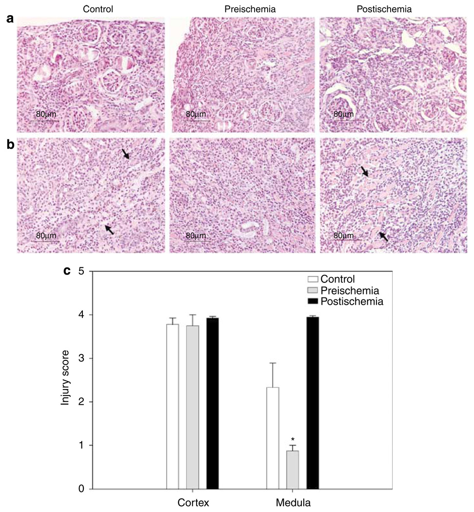
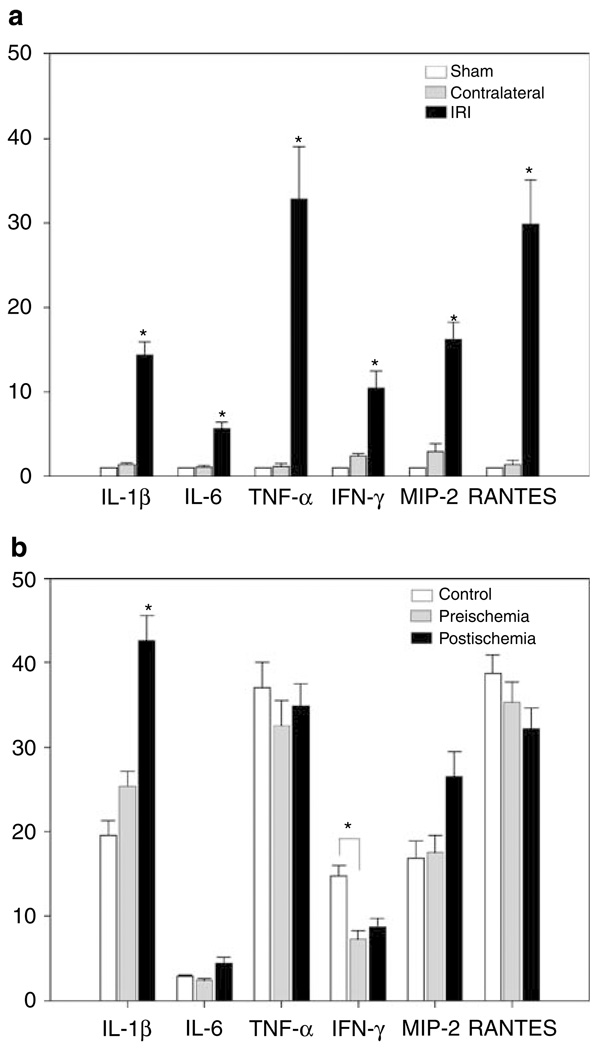
It is well-established that significant ischemia-reperfusion injury during kidney transplantation results in increased incidence of long-term fibrosis and rejection. To test for a role of T cell infiltration and activation following ischemic injury, we induced both bilateral and unilateral renal ischemia in mice, followed by reperfusion, and then isolated mononuclear cells. Analysis of these cells by flow cytometry showed that 2 weeks after bilateral ischemia there was a significant increase of CD8(+) T cells. Furthermore, both CD4(+) and CD8(+) T cells infiltrated the injured kidney 6 weeks after unilateral ischemia. These T cells had increased expression of CD69(+) and CD44(hi)CD62L(-), markers of activation and effector-memory, respectively. CD4(+)NK1.1(+) and CD19(+) B cells were decreased in percentage both 6 and 11 weeks after bilateral or unilateral injury. There was a significant upregulation of IL-1beta, IL-6, TNF-alpha, IFN-gamma, MIP-2, and RANTES expression, measured by real-time PCR, 6 weeks after unilateral renal ischemia, further indicating T cell activation. Depletion of CD4(+) and CD8(+) T cells before ischemia caused less medullary damage and reduced kidney IFN-gamma expression, whereas their depletion following ischemia increased kidney IL-1beta; however, depletion of these cells had no effect on histological damage to the kidney. Our study demonstrates that moderate or severe kidney ischemia induces long-term T lymphocyte infiltration and cytokine/chemokine upregulation, leading to kidney structural changes.


Figures

Figure 1
Figure 1. Mouse kidney tissue injury 2 weeks after bilateral and 6 weeks after unilateral renal IRI
Kidney histology in tissue sections stained with H&E was observed at original magnification × 200. Kidney tissue from IRI mice after 2 weeks of 25 min of bilateral ischemia (a, upper panel) shows some proteinaceous casts in tubules compared with normal histology of normal and sham mouse kidneys. Kidney structure 6 weeks after unilateral renal IRI (b, lower panel) shows normal histology of sham and contralateral kidneys compared with severe kidney damage, loss of structure, and cyst formation in IRI kidneys.
Figure 2
Figure 2. Increased expression of CD69 + activation marker on CD4 + and CD8 + T-cell subsets long term after ischemic injury
Freshly isolated KMNC were stained with mAb anti-CD8 + FITC, anti-CD4 + PerCP, and anti-CD69 + PE and analyzed by flow cytometry. Increased expression of early activation marker CD69 on CD8 + T cells after 2 (*P = 0.001) and 6 (*P = 0.033) weeks of bilateral renal IRI (a, left panel) was observed. After 6 weeks of unilateral ischemia, both CD4 + (*P < 0.001) and CD8 + (*P = 0.013) T cells increased CD69 expression. Increased percentage of CD4 + CD69 + T cells 11 weeks after-unilateral IRI (b, right panel) was also observed. Cells were gated in the lymphocyte and CD3 + T-cell areas and 10,000 events were collected. Values of the bar graphs represent the percentage mean ± s.e.m. (n = 3 to 4 per group.)
Figure 3
Figure 3. Increased expression of effector-memory CD4 + CD44 hiCD62L T-cell phenotype after unilateral and bilateral renal IRI
The percentages of positive cells were obtained from the gated lymphocyte, CD3 + and CD4 + (R3) T-cell areas. Increased percentage of CD4 + CD44hiCD62L T cells in IRI kidneys (*P = 0.001) compared with kidneys from sham mice after 2 weeks of bilateral renal IRI is shown (a, upper panel). Increased percentage of CD4 + CD44hiCD62L T cells 6 weeks after unilateral treatment (*P < 0.001; b, lower panel) is shown. Representative dot plots show the mean percentage of positive cells ± s.e.m. (n = 3 to 4 per group.)
Figure 4
Figure 4. Decreased percentages of CD4 +NK1.1 + cells and CD19 + B cells after bilateral and unilateral renal IRI
Decreased percentage of CD4 + NK1.1 + T cells is shown in IRI kidneys after 6 weeks of bilateral (*P = 0.007) and 11 weeks of unilateral (*P = 0.004) renal IRI when compared with control kidneys (a, left panel). The percentage of CD19 + B cells also decreased significantly in IRI kidneys after 6 (*P = 0.004) and 11 (*P < 0.001) weeks of unilateral renal IRI (b, right panel). Values of the bar graphs represent the percentage mean ± s.e.m. (n = 3 to 4 per group.)
Figure 5
Figure 5. Effect of CD4 + and CD8 + T-cell depletion 6 weeks after severe ischemia
Depletion of CD4 + and CD8 + T cells in peripheral blood was around 98% during the 6 weeks after ischemic insult (a, upper panel), in comparison to control nondepleted mice. Depletion of CD4 + CD69 +, CD8 + CD69 +, CD4 + CD44hiCD62L, and CD4 + NK1.1 + T cells in kidneys was around 98%, in comparison to control nondepleted mice (b, lower panel). Representative dot plot examples show the mean percentage of positive cells ± s.e.m. (n = 4 per group.)
Figure 6
Figure 6. Decreased percentages of CD19 + B cells after unilateral renal IRI in both depleted and control (nondepleted) mice
CD19 + B cells were plotted with CD4 + T cells to observe the difference between depleted and control nondepleted mice. No significant difference existed between control and depleted mice. However, the percentage of CD19 + B cells decreased significantly in IRI kidneys in both control (*P < 0001; a, upper panel) and depleted (*P = 0.028; b, lower panel) mice, after 6 weeks of unilateral renal IRI. Values of the bar graph (c, right panel) represent the percentage of B cells in control and depleted mice (mean ± s.e.m., n = 4 per group).
Figure 7
Figure 7. Histology assessment of kidney tissue sections stained with H&E
Kidney structure in depleted and control (nondepleted) mice 6 weeks after unilateral ischemia demonstrates damage, loss of structure and cyst formation in IRI kidneys. Pictures show the cortex (a, upper panel) and medulla (b, lower panel) damage to control and both (preischemia and postischemia) depleted mice. Injury score evaluation of kidneys (c), shows same damage in cortex of control and both depleted mice, but lower damage (*P = 0.029) in medulla of preischemia depleted mice (mean ± s.e.m., n = 4 to 8 per group). Original magnification was × 200.
Figure 8
Figure 8. RT–PCR analysis of kidney cytokines and chemokines from CD4 + and CD8 + T-cell depleted and control mice
Total RNA was isolated from mouse kidneys 6 weeks after 60 min unilateral renal IRI. Relative gene expressions of IL-1β, IL-6, TNF-α, IFN-γ, MIP-2, and RANTES are displayed as fold change values presented as average fold change 2−ΔΔCt method, between IRI mice and control kidneys. High expression (*P < 0.05) of all cytokine and chemokine factors is observed in IRI kidneys of normal nondepleted mice when compared with sham and contralateral ones (a, upper panel). A significant (*P < 0.05) decrease of IFN-γ in preischemia depleted mice, and a significant (*P < 0.05) increase of IL-1β in postischemia mice were observed when compared to profiles of control mice (b, lower panel). Bar graphs represent the percentage mean ± s.e.m. (n = 4 to 8 per group.)

References

    1. Tilney NL, Guttmann RD. Effects of initial ischemia/reperfusion injury on the transplanted kidney. Transplantation. 1997;64:945–947. - PubMed
    1. Humes HD. Acute renal failure: prevailing challenges and prospects for the future. Kidney Int. 1995;50:S26–S32. - PubMed
    1. Terasaki PI, Cecka JM, Gjertson DW, et al. survival rates of kidney transplants from spousal and living unrelated donors. N Engl J Med. 1995;333:333–336. - PubMed
    1. van Es A, Hermans J, van Bockel JH, et al. Effect of warm ischemia time and HLA (A and B) matching on renal cadaveric graft survival and rejection episodes. Transplantation. 1983;36:255–258. - PubMed
    1. Sanfilippo F, Vaughn WK, Spees EK, et al. The detrimental effects of delayed graft function in cadaver donor renal transplantation. Transplantation. 1984;38:643–648. - PubMed

Publication types