. 2008;3(12):e3917.
doi: 10.1371/journal.pone.0003917. Epub 2008 Dec 18.

Olig2-induced neural stem cell differentiation involves downregulation of Wnt signaling and induction of Dickkopf-1 expression

Affiliations

Olig2-induced neural stem cell differentiation involves downregulation of Wnt signaling and induction of Dickkopf-1 expression

Sung-Min Ahn et al. PLoS One. 2008.

Abstract

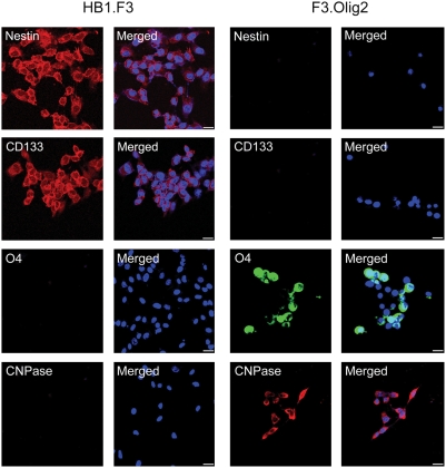
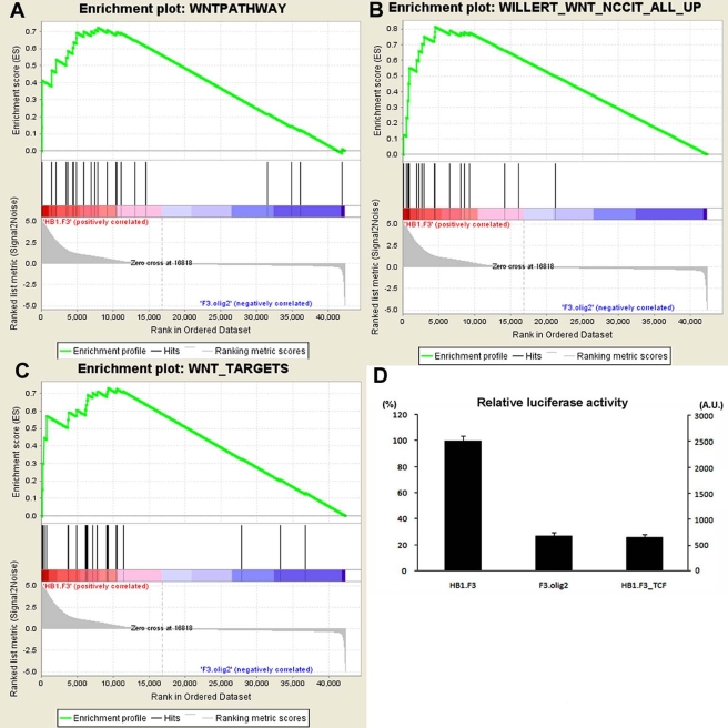
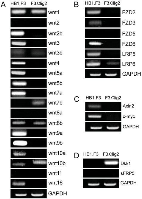
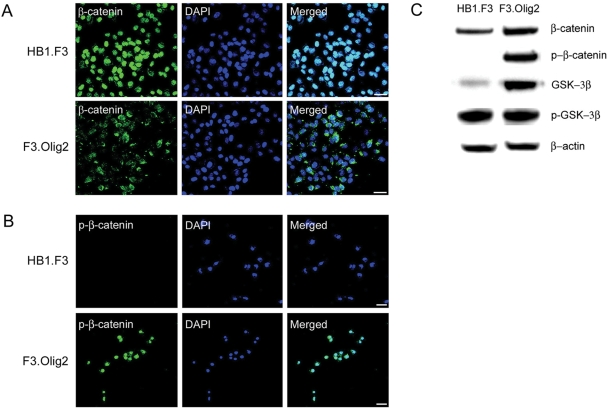
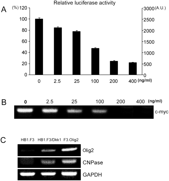
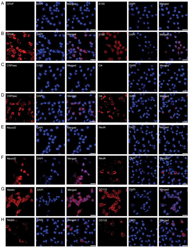
Understanding stem cell-differentiation at the molecular level is important for clinical applications of stem cells and for finding new therapeutic approaches in the context of cancer stem cells. To investigate genome-wide changes involved in differentiation, we have used immortalized neural stem cell (NSC) line (HB1.F3) and Olig2-induced NSC differentiation model (F3.Olig2). Using microarray analysis, we revealed that Olig2-induced NSC differentiation involves downregulation of Wnt pathway, which was further confirmed by TOPflash/FOPflash reporter assay, RT-PCR analysis, immunoblots, and immunocytochemistry. Furthermore, we found that Olig2-induced differentiation induces the expression of Dickkopf-1(Dkk1), a potent antagonist of Wnt signaling. Dkk1 treatment blocked Wnt signaling in HB1.F3 in a dosage-dependent manner, and induced differentiation into astrocytes, oligodendrocytes, and neurons. Our results support cancer stem cell hypothesis which implies that signaling pathway for self-renewal and proliferation of stem cells is maintained till the late stage of differentiation. In our proposed model, Dkk1 may play an important role in downregulating self-renewal and proliferation pathway of stem cells at the late stage of differentiation, and its failure may lead to carcinogenesis.


Conflict of interest statement

Competing Interests: The authors have declared that no competing interests exist.

Figures

Figure 1
Figure 1. Expression of lineage-specific markers in HB1.F3 and F3.Olig2.
Neural stem cell markers such as nestin and CD133 are expressed only in HB1.F3 wherease oligodendrocytes markers such as O4 and CNPase are expressed only in F3.Olig2. Merged; markers with DAPI, Bar = 50 µm
Figure 2
Figure 2. Downregulation of Wnt pathway in F3.Olig2.
GSEA shows that Wnt pathway genes (A), Wnt-responsive genes (B), and Wnt pathway target genes (C) are enriched in HB1.F3. TOPFLASH/FOPflash reporter analysis (D) confirms that Wnt pathway activity in HB1.F3 is significantly higher than that in F3.Olig2. The reporter activity in HB1.F3 was inhibited to the level of F3.Olig2 when HB1.F3 was co-transfected with ‘dominant negative’ (DN) TCF constructs. Data are means±S.D. of triplicate samples. A.U., Activity unit
Figure 3
Figure 3. RT-PCR analysis of Wnt pathway-related genes.
A. Most Wnt genes are overexpressed in HB1.F3 whereas the expression of Wnt 10b is increased in F3.Olig2. Interestingly, Wnt7b is expressed only in F3.Olig2. B. Except FZD5, all Wnt receptor and co-receptor genes that are expressed in HB1.F3 are suppressed in F3.Olig2. C. Wnt pathway target genes are expressed only in HB1.F3. D. Dkk1, a soluble Wnt antagonist, is expressed only in F3.Olig2.
Figure 4
Figure 4. Levels of β-catenin and phospho-β-catenin (p-β-catenin), and their subcellular localization.
In HB1.F3, β-catenin is mainly localized in nucleus (A), and p-β-catenin is not detected (B, C). In F3.Olig2, β-catenin is mainly localized in cytoplasm (A), and p-β-catenin is detected and mainly localized in nucleus (B, C). The level of GSK3β, which phosphorylates β-catenin on Ser-33/Ser-37/Thr-41, is increased in F3.Olig2 (C). Bar = 50 µm
Figure 5
Figure 5. Dkk1 inhibits Wnt signaling in HB1.F3.
A. TOPflash/FOPflash reporter assay shows that Dkk1 represses Wnt signaling in HB1.F3 in a dosage-dependent manner. Data are means±S.D. of triplicate samples. B. Dkk1 represses the expression of c-myc, one of the target genes of Wnt pathway, in HB1.F3 in a dosage-dependent manner. C. Dkk1 treatment induces the expression of oligodendrocytes markers (Olig2 and CNPase) in HB1.F3.
Figure 6
Figure 6. Immunohistochemical staining of HB1.F3 or HB1.F3 with Dkk1 treatment for various markers of stem cell and differentiated cells.
Dkk1 induces differentiation of HB1.F3 into astrocytes, neurons and oligodendrocytes. Astrocyte markers (GFAP, S100), oligodendrocyte markers (CNPase, O4), neuron markers (NeuroD, NeuN) and neural stem cell markers(Nestin, CD133) were double stained with DAPI (blue) in HB1.F3 before (A, C, E, G ) and after (B, D, F, H ) Dkk1 treatment. Bar = 50 µm

Similar articles

Cited by

References

    1. Kim SU. Human neural stem cells genetically modified for brain repair in neurological disorders. Neuropathology. 2004;24:159–171. - PubMed
    1. Cattaneo E, McKay R. Proliferation and differentiation of neuronal stem cells regulated by nerve growth factor. Nature. 1990;347:762–765. - PubMed
    1. Kilpatrick TJ, Bartlett PF. Cloning and growth of multipotential neural precursors: requirements for proliferation and differentiation. Neuron. 1993;10:255–265. - PubMed
    1. Temple S. Division and differentiation of isolated CNS blast cells in microculture. Nature. 1989;340:471–473. - PubMed
    1. Lois C, Alvarez-Buylla A. Proliferating subventricular zone cells in the adult mammalian forebrain can differentiate into neurons and glia. Proc Natl Acad Sci U S A. 1993;90:2074–2077. - PMC - PubMed

Publication types

MeSH terms

Substances