. 2008 Nov-Dec;1(4-5):229-40.
doi: 10.1242/dmm.000729. Epub 2008 Nov 6.

Peptidylarginine deiminase 2 (PAD2) overexpression in transgenic mice leads to myelin loss in the central nervous system

Affiliations

Peptidylarginine deiminase 2 (PAD2) overexpression in transgenic mice leads to myelin loss in the central nervous system

Abdiwahab A Musse et al. Dis Model Mech. 2008 Nov-Dec.

Abstract

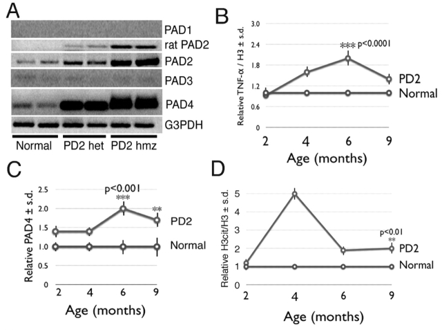
Demyelination in the central nervous system is the hallmark feature in multiple sclerosis (MS). The mechanism resulting in destabilization of myelin is a complex multi-faceted process, part of which involves deimination of myelin basic protein (MBP). Deimination, the conversion of protein-bound arginine to citrulline, is mediated by the peptidylarginine deiminase (PAD) family of enzymes, of which the PAD2 and PAD4 isoforms are present in myelin. To test the hypothesis that PAD contributes to destabilization of myelin in MS, we developed a transgenic mouse line (PD2) containing multiple copies of the cDNA encoding PAD2, under the control of the MBP promoter. Using previously established criteria, clinical signs were more severe in PD2 mice than in their normal littermates. The increase in PAD2 expression and activity in white matter was demonstrated by immunohistochemistry, reverse transcriptase-PCR, enzyme activity assays, and increased deimination of MBP. Light and electron microscopy revealed more severe focal demyelination and thinner myelin in the PD2 homozygous mice compared with heterozygous PD2 mice. Quantitation of the disease-associated molecules GFAP and CD68, as measured by immunoslot blots, were indicative of astrocytosis and macrophage activation. Concurrently, elevated levels of the pro-inflammatory cytokine TNF-alpha and nuclear histone deimination support initiation of demyelination by increased PAD activity. These data support the hypothesis that elevated PAD levels in white matter represents an early change that precedes demyelination.


Figures

Fig. 1
Fig. 1
PAD2 construct and clinical signs in PAD2 transgenic mice. (A) The rat PAD2 transgene construct. (B) Southern blot analysis of rat PAD2 transgene expression in mouse line numbers 28, 25, 15 and 9, with endogenous PLP used as an internal standard. The ‘+/–’ designation indicates transgene expression in the heterozygotes, whereas ‘–/–’ indicates the absence of the transgene. (C) Progression of clinical disease in PD2 mice (n=10) relative to ND4 mice (n=10). Clinical scores comparing the observed neurological signs of the spontaneously demyelinating ND4 mice with those of PD2 mice are shown. The measured neurological signs included tail droop, tremors, unsteady gait, head shaking, convulsions, physical activity, and righting ability (a measure of strength). The mean clinical signs for normal non-transgenic littermates fell within the range of 0–10, which is represented by the shaded area. The values shown are the aggregated means of the neurological signs. The scores ranged from 0 (no signs) to 40 (severe).
Fig. 2
Fig. 2
PAD2 levels are elevated in PD2 white matter and in brain homogenates from PD2 mice. (A) Immunohistochemical analysis showing the extent of PAD2 expression in normal and heterozygous PD2 mice using anti-PAD2 antibody. Representative corpus callosum regions (arrows) of paraffin-embedded, formalin-fixed, sections of brain from 6-month-old normal control and heterozygous PD2 mice were probed with anti-pan-PAD2 antibody and counterstained with hematoxylin. (B) Immunoslot blot showing quantitative expression levels of PAD2, normalized to histone H3, in normal and heterozygous PD2 mice, at various ages, using anti-PAD2 antibody and anti-histone H3 antibody (loading control). Significantly increased PAD2 levels are found at 2 (P<0.02), 4 (P<0.002) and 6 (P<0.002) months of age. (C) Specific PAD activity in isolated myelin from normal and heterozygous transgenic PD2 mice of different ages. All measurements represent the means ± s.d. of at least 10 independent determinations. Significant differences in specific PAD activity were observed in the myelin isolated from heterozygous PD2 transgenic mice compared with normal (N) mice at 1 (P<0.01), and 2 (P<0.0002) months of age. (D) Immunoslot blot analysis of the extent of MBP citrullination in isolated myelin from normal (N) and PD2 mice. The normalized relative ratios of deiminated and total MBP, determined using anti-modified citrulline antibody and anti-MBP antibody, respectively, are shown. The error bars indicate the s.d. from 9 determinations of each sample. Significant differences were observed at 2 (P<0.02) and 3 (P<0.0002) months of age, consistent with the elevated levels of PAD2 activity.
Fig. 3
Fig. 3
White matter changes are revealed by morphological analysis in PD2 mice. (A-C) Light microscopic analysis of Toluidine Blue-stained semithin epon-embedded cross-sections of optic nerves from 6-month-old non-transgenic normal mice (A), and heterozygous (B) and homozygous (C) PD2 transgenic mice. Although subtle, mild changes in axon thickness were detected in the heterozygous PD2 transgenic mice. Note the decrease in number and diameter of the myelinated axons in the homozygous PD2 mice. In addition, evidence of modest trabeculation (arrowheads) consistent with an atropic nerve is evident. Image width of A, B and C is 0.1 mm. (D–E) LFB-Holmes-stained histological sections of non-transgenic (D) and homozygous PD2 (E) cerebellar folia revealed a higher amount of coarse fibers in the homozygous PD2 mice. White matter (WM) is stained blue. The gray matter (GM) granular layer is stained pink. Bar, 1 μm.
Fig. 4
Fig. 4
Morphological changes in myelinated fibers from heterozygous PD2 mice revealed by transmission electron microscopy. (A) Electron micrographs of optic nerve cross-sections from a non-transgenic mouse. Myelinating axons are seen in close proximity to oligodendrocyte processes. (B) Heterozygous PD2 optic nerve cross-section reveals a normally myelinated area. (C) Heterozygous PD2 optic nerve cross-section reveals thinly myelinated fibers (arrows) and nude axons (arrowhead), and an astrocyte process in the lower left corner of the micrograph. Bars, 1 μm.
Fig. 5
Fig. 5
Demyelination in PD2 homozygous mice. Low power transmission electron micrograph of a cross-section from a homozygous PD2 optic nerve (A) reveals myelinated axons and astrocytic processes (asterisks), which are seen between the axons, denuded axons and myelin debris. (B) Higher power micrograph of (A) showing that degenerating (arrow) and nude axons (arrowheads) were observed in many areas containing astrocytic processes (asterisks). In areas of variably myelinated axons in optic nerves from homozygous PD2 mice (C,D), denuded axons (arrowheads) and astrocytic processes (asterisk) are seen. Bars, 1 μm.
Fig. 6
Fig. 6
Lymphocyte infiltration and astrocytosis in PD2 mice. (A) CD3, CD8 and CD68 levels in the brains of PD2 mice. Immunoslot blots were used to determine the reactivity of CD3, CD8 and CD68, using anti-CD3, anti-CD8 and anti-CD68 antibodies, in brain homogenates from SJL mice with acute EAE, and from 2-, 4-, 6- and 9-month-old normal and heterozygous PD2 mice. The loading control for each individual was reactivity of the anti-histone H3 antibody. (B) Immunoslot blot analysis of the amount of GFAP in normal and heterozygous PD2 mice, as measured by GFAP immunoreactivity in whole brain homogenates using anti-GFAP antibody. The graph shows the amount of GFAP in PD2 mice relative to their normal littermates, as determined from the ratio of GFAP to the loading standard mouse actin. At each age, the ratios of GFAP to actin pixel densities from the PD2 mice were normalized to those of their normal littermates.
Fig. 7
Fig. 7
PAD2 and PAD4 mRNA is elevated in PD2 white matter. (A) PAD cDNAs (specific for PAD 1, 2, 3 and 4) from RT-PCR of the total cDNA isolated from mouse brain white matter, separated by agarose gel electrophoresis. PAD1-specific primers revealed no amplicons. PAD3-specific primers revealed PCR products, but their abundance was not significant and was much lower than the levels in mouse skin control cDNA (data not shown). (B) Relative amounts of TNF-α in the brains of PD2 mice at 2, 4, 6 and 9 months of age. The amount of TNF-α at each age was normalized against the amount of histone H3 in brain extracts. Each data point represents the average ± s.d. of 6 measurements. (C) Relative amounts of PAD4 in nuclear fractions prepared from the brains of normal and PD2 mice at 2, 4, 6 and 9 months of age. Nuclear fractions were prepared as we described previously (Mastronardi et al., 2006). (D) Relative amounts of citrullinated histone H3 in PD2 brains at 2, 4, 6 and 9 months of age. The amount of citrullinated histone H3 at each age was normalized against the amount of total histone H3 in brain extracts. Each data point represents the average ± s.d. of 3 measurements.
Fig. 8
Fig. 8
Model of disease pathogenesis. The hypomethylation of genomic DNA was found to be associated with increased DNA demethylase activity, which was found to be 2′ higher in MS white matter compared with controls (Mastronardi et al., 2007a). Hypomethylation of the PAD2 promoter results in increased expression of PAD2 in myelin, leading to increased MBP citrullination and subsequently unstable myelin. Formation of citrullinated MBP at the myelin periaxonal structures may result in an early cascade of events resulting in oligodendrocyte apoptosis. Myelin instability also results in the release of immunogenic epitopes. These epitopes include MBP peptides capable of stimulating the immune response. The immune response feeds forward with further demyelination and more damage. Extrinsic signals, such as TNF-α, mobilize PAD4 enzyme into the nucleus, where it deiminates histones (Mastronardi et al., 2006). Mobilization of PAD4 into the nucleus may be an additional mechanism that contributes to oligodendrocyte apoptosis.

Similar articles

Cited by

References

    1. Akassoglou K., Bauer J., Kassiotis G., Pasparakis M., Lassmann H., Kollias G., Probert L. (1998)Oligodendrocyte apoptosis and primary demyelination induced by local TNF/p55TNF receptor signaling in the central nervous system of transgenic mice: models for multiple sclerosis with primary oligodendrogliopathy. Am. J. Pathol. 153, 801–813 - PMC - PubMed
    1. Akassoglou K., Bauer J., Kassiotis G., Lassmann H., Kollias G., Probert L. (1999)Transgenic models of TNF induced demyelination. Adv. Exp. Med. Biol. 468, 245–259 - PubMed
    1. Bruck W. (2007)New Insights into the pathology of multiple sclerosis: towards a unified concept. J. Neurol. 254, 3–9
    1. Chaudhuri A., Behan P. O. (2005)Multiple sclerosis: looking beyond autoimmunity. J. R. Soc. Med. 98, 303–306 - PMC - PubMed
    1. Chung I. Y., Benveniste E. N. (1990)Tumor necrosis factor-alpha production by astrocytes: induction by lipopolysaccharide, IFN-gamma, and IL-1 beta. J. Immunol. 144, 2999–3007 - PubMed

Publication types