. 2008 Nov-Dec;1(4-5):241-54.
doi: 10.1242/dmm.000711. Epub 2008 Nov 6.

Analysis of mouse models carrying the I26T and R160C substitutions in the transcriptional repressor HESX1 as models for septo-optic dysplasia and hypopituitarism

Affiliations

Analysis of mouse models carrying the I26T and R160C substitutions in the transcriptional repressor HESX1 as models for septo-optic dysplasia and hypopituitarism

Ezat Sajedi et al. Dis Model Mech. 2008 Nov-Dec.

Abstract

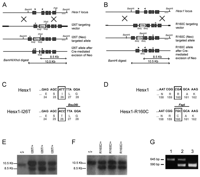
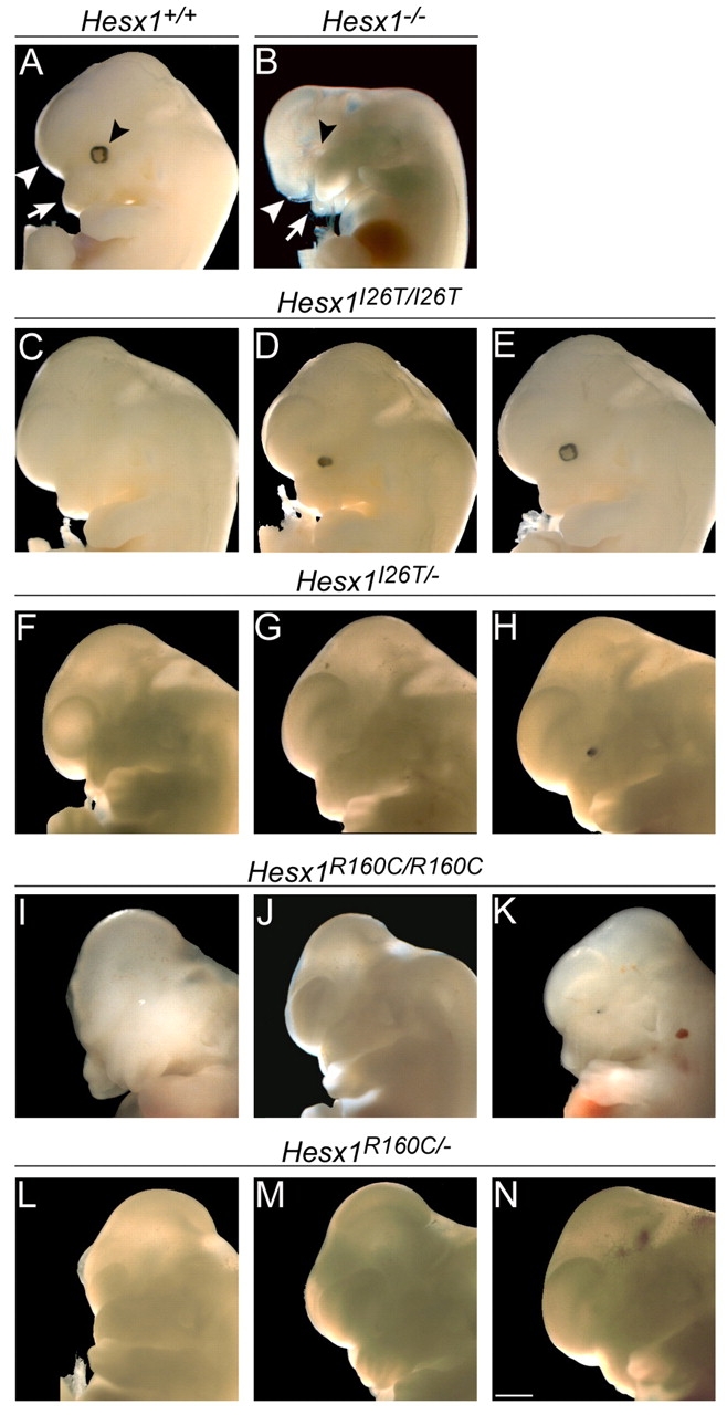
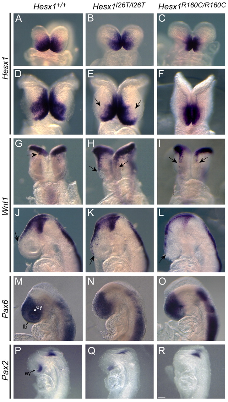
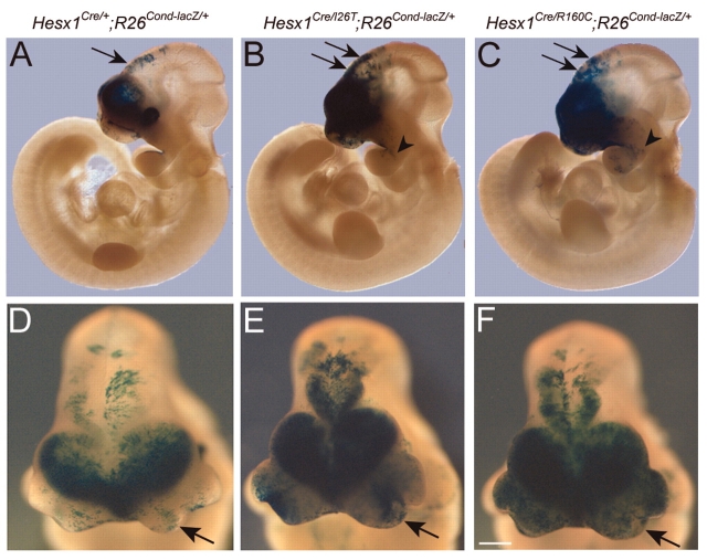
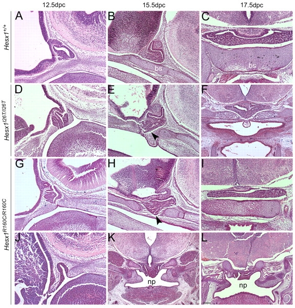
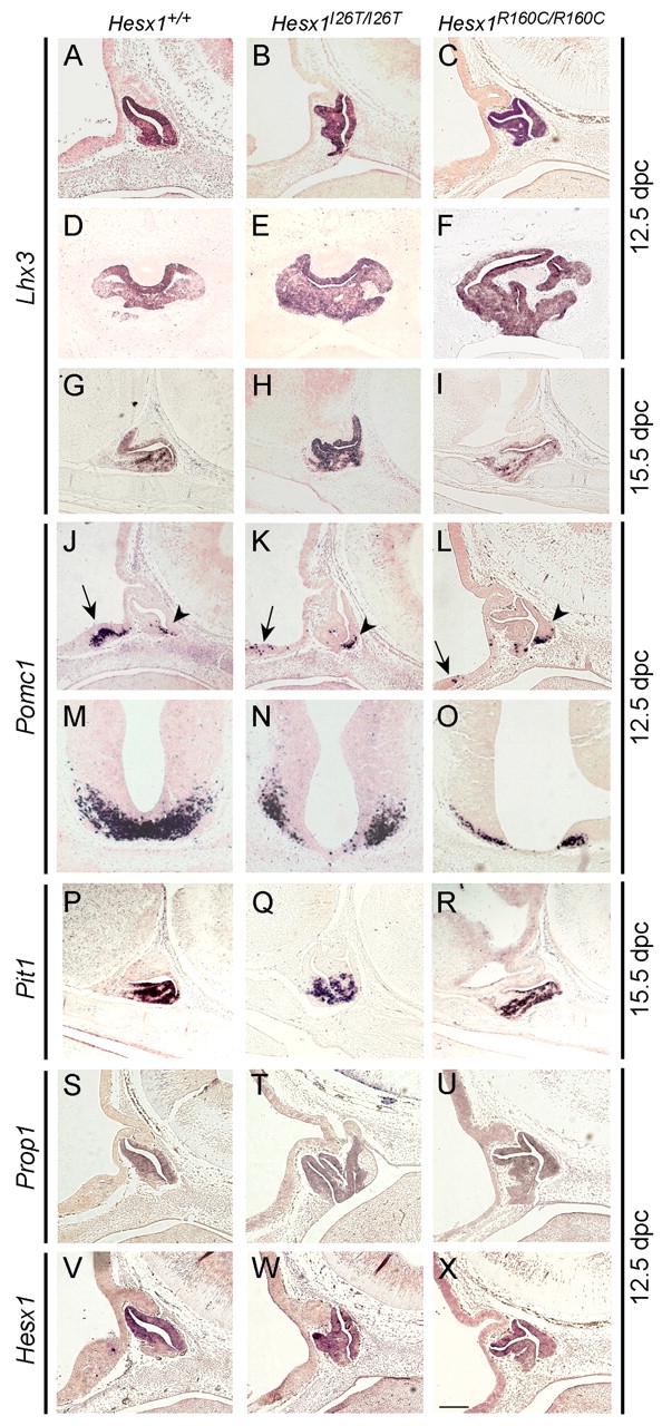
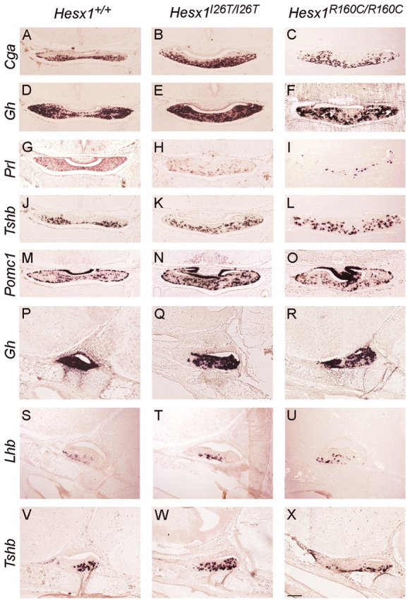
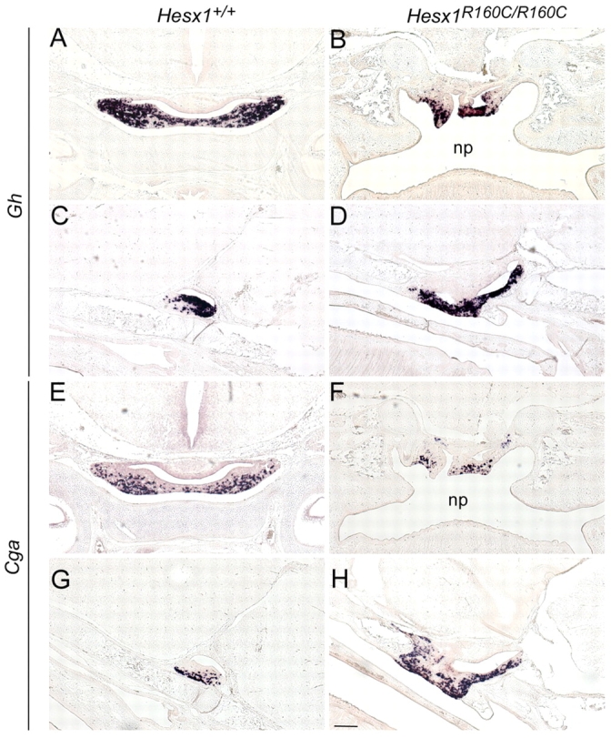
A homozygous substitution of the highly conserved isoleucine at position 26 by threonine (I26T) in the transcriptional repressor HESX1 has been associated with anterior pituitary hypoplasia in a human patient, with no forebrain or eye defects. Two individuals carrying a homozygous substitution of the conserved arginine at position 160 by cysteine (R160C) manifest septo-optic dysplasia (SOD), a condition characterised by pituitary abnormalities associated with midline telencephalic structure defects and optic nerve hypoplasia. We have generated two knock-in mouse models containing either the I26T or R160C substitution in the genomic locus. Hesx1(I26T/I26T) embryos show pituitary defects comparable with Hesx1(-/-) mouse mutants, with frequent occurrence of ocular abnormalities, although the telencephalon develops normally. Hesx1(R160C/R160C) mutants display forebrain and pituitary defects that are identical to those observed in Hesx1(-/-) null mice. We also show that the expression pattern of HESX1 during early human development is very similar to that described in the mouse, suggesting that the function of HESX1 is conserved between the two species. Together, these results suggest that the I26T mutation yields a hypomorphic allele, whereas R160C produces a null allele and, consequently, a more severe phenotype in both mice and humans.


Figures

Fig. 1
Fig. 1
Generation of the Hesx1-I26T and Hesx1-R160C targeted alleles. (A,B) Top to bottom: structure of the murine Hesx1 locus; Hesx1-I26T (A) and Hesx1-R160C (B) targeting vectors; targeted alleles prior to and after Cre-mediated excision of the Neo cassette; expected bands for the targeted and wild-type alleles after Southern blot analysis of DNA samples digested with the indicated restriction enzymes and hybridised with an external probe (dotted line). The position of the mutation is indicated with an asterisk on exons one (I26T) and four (R160C) of the targeting vectors and targeted alleles. Note that the orientation of the loxP and Neo cassette has been inverted in the Hesx1-R160C targeting vector. (C) The triplet ATT encoding isoleucine at position 26 was replaced by ACC, which encodes the amino acid threonine. This mutation introduces a novel Bsu36I restriction site on the mutated allele. (D) The triplet encoding arginine at position 160 was replaced by TGC, which encodes the amino acid cysteine. This mutation introduces a novel FspI restriction site in the mutated allele. (E,F) Southern blot analysis of DNA samples from wild-type (+/+), Hesx1I26T/+ (E) and Hesx1R160C/+ (F) ES cell clones digested with either BamHI/XhoI (E) or BamHI (F) and hybridised with an external probe (dotted line in A,B). (G) Representative example of PCR genotyping of DNA samples from the homozygous Hesx1I26T/I26T or Hesx1R160C/R160C (1), heterozygous Hesx1I26T/+ or Hesx1 R160C/+ (2) and wild-type Hesx1+/+ (3) embryos. Note that the primers used for genotyping both targeted alleles are the same [black arrowheads in (A) and (B)].
Fig. 2
Fig. 2
Forebrain defects in mutant embryos harbouring the Hesx1-I26T or Hesx1-R160C alleles. Dark-field photographs of 12.5 dpc embryos of specific genotypes (indicated on the top of the pictures). (A,B) Wild-type (A) and Hesx1–/– (B) embryos. Hesx1–/– embryos carry a null allele, in which a Neo cassette replaces the entire Hesx1 coding region. Note the absence of eyes (black arrowhead), reduced telencephalic vesicles (white arrowhead) and impairment of frontonasal mass development (white arrow) in the Hesx1–/–mutant (B) when compared with the wild-type embryo (A). (C-E) Representative examples of Hesx1I26T/I26T mutants displaying anophthalmia (C), microphthalmia (D) or normal eyes (E). Telencephalic vesicles are unaffected. (F-H) Representative examples of Hesx1I26T/– mutants. Note the increased severity in the eye and telencephalic defects in these embryos when compared with Hesx1I26T/I26T embryos (C-E). (I-K) Representative examples of Hesx1R160C/R160C mutants showing severe forebrain defects (I) and either anophthalmia (J) or microphthalmia (K) with normal telencephalic vesicles. (L-M) Representative examples of Hesx1R160C/– embryos. There is no obvious increase in the severity of eye and telencephalic abnormalities between embryos carrying one (L-M) or two (I-K) copies of the Hesx1-R160C allele. Bar, 940 μm.
Fig. 3
Fig. 3
Early forebrain patterning defects in Hesx1I26T/I26T and Hesx1R160C/R160C embryos. Photographs of whole-mount in situ hybridisations. Probes used are indicated on the left side. (A-F) Frontal views of 8.0 dpc (A-C) and 8.5 dpc (D-F) embryos hybridised with Hesx1 antisense riboprobes. The expression domain of Hesx1 is reduced in Hesx1I26T/I26T embryos (B,E) when compared with wild-type embryos (A,D). Note that at 8.5 dpc the reduction is apparent in the proximal region of the developing optic cups (arrows in E). Reduction of the Hesx1 expression domain is more accentuated in Hesx1R160C/R160C embryos (C,F). (G-L) Frontal (G-I) and lateral (J-L) views of embryos hybridised with Wnt1 antisense riboprobes at 8.5 and 9.0 dpc. Note the anteriorisation of the Wnt1 expression domain in Hesx1I26T/I26T (H,K) and Hesx1R160C/R160C (I,L) mutants when compared with wild-type embryos (G,J). The arrows in G-L indicate the rostral limit of the Wnt1 expression domain in the dorsal forebrain. (M-O) Lateral views of embryos hybridised with Pax6 antisense riboprobes at 9.0 dpc. The Pax6 expression domain is reduced in the Hesx1I26T/I26T mutants (N) as the eyes (ey) are smaller in size (see text for details). Hesx1R160C/R160C mutants (O) show a more severe reduction in forebrain tissue (fb) and no eyes compared with Hesx1I26T/I26T mutants (N). (P-R) Lateral views of embryos hybridised with Pax2 antisense riboprobes at 8.5 dpc. Pax2 is expressed in the eye (ey) and the mid-hindbrain boundary in a wild-type embryo (P). Note that reduction or absence of eye tissue in the Hesx1I26T/I26T (Q) or Hesx1R160C/R160C (R) mutants is concomitant with a reduction in Pax2 expression in the eye. Bar: 130 μm (A-C,J-O), 170 μm (D-F), 215 μm (G-I), 40 μm (P-R).
Fig. 4
Fig. 4
Posterior transformation of anterior forebrain precursors in Hesx1Cre/I26T and Hesx1Cre/R160C mutants. (A-F) X-Gal staining on 10.0 dpc embryos of the three genotypes (indicated on the top of the columns) reveals abundant lacZ-positive cells in the posterior forebrain (arrowheads) of the Hesx1Cre/I26T;R26Cond-lacZ/+ (B,E) and Hesx1Cre/R160C;R26Cond-lacZ/+ (C,F) mutants in comparison with the Hesx1Cre/+;R26Cond-lacZ/+ control embryo (A,D). Also note that lacZ-positive cells are present within the first branchial arch of the mutant embryos (arrows in B,C), whereas, in the control embryo (A), they colonise only the endodermal lining of the first branchial arch. Bar: 550 μm (A-C), 180 μm (D-F).
Fig. 5
Fig. 5
Histological analysis of the developing pituitary gland in Hesx1I26T/I26T and Hesx1R160C/R160C mutants. H&E staining of embryos (genotypes are indicated on the left side of the panel) at different stages of development (indicated on top of the panel). (A-C) Wild-type pituitary gland. (D-I) Hesx1I26T/I26T (D-F) and Hesx1R160C/R160C (G-I) pituitary glands showing the type I phenotype, which typically display anterior pituitary enlargement and bifurcation with or without defective development of the basisphenoid cartilage (bs). (J-L) Hesx1R160C/R160C pituitary gland displaying the type II phenotype, which is characterised by a delay of Rathke’s pouch development at 12.5 dpc (J), severe impairment of basisphenoid cartilage development and ectopic pituitary in the nasopharynx (np) (K,L). Bar, 110 μm.
Fig. 6
Fig. 6
Rathke’s pouch is dysmorphic but properly specified in Hesx1I26T/I26T and Hesx1R160C/R160C mutants. In situ hybridisation (ISH) on sagittal (A-C, G-L and P-X) and frontal (D-F and M-O) paraffin sections of embryos of different genotypes (indicated on the top of each column). The probes used for ISH and the developmental stages of embryos analysed are indicated on the left and right sides of the panel, respectively. (A-I) Lhx3 expression is normal, but Rathke’s pouch is expanded showing aberrant morphology and contains bifurcated lumens in the Hesx1I26T/I26T (B,E,H) and Hesx1R160C/R160C (C,F,I) mutants when compared with wild-type embryos (A,D,G). (J-L) Pomc1 expression in Rathke’s pouch is not affected (arrowheads), but there is reduced Pomc1 expression in the hypothalamic area (arrows) of the Hesx1I26T/I26T (K) and Hesx1R160C/R160C (L) embryos in comparison with the wild-type embryo (J). (M-O) Reduction of Pomc1 expression in the hypothalamic area is apparent in frontal sections. (P-R) Pit1 expression in ventral progenitors is indistinguishable between genotypes. (S-U) Prop1 expression is unaffected in Hesx1I26T/I26T (T) and Hesx1R160C/R160C (U) embryos, but Rathke’s pouch morphology is aberrant. (V-X) Hesx1 transcripts are detected in the developing Rathke’s pouch in a high-dorsal to low-ventral gradient of expression in the three genotypes. Bar: 155 μm (A-L,P-X), 100 μm (M-O).
Fig. 7
Fig. 7
Terminal differentiation of hormone-producing cells occurs normally in the anterior pituitary of Hesx1I26T/I26T and Hesx1R160C/R160C embryos. In situ hybridisation (ISH) on frontal (A-O) or sagittal (P-X) paraffin sections of 17.5 dpc embryos. Genotypes and probes used for ISH are indicated on the top and left sides of the panel, respectively. (A-X) The levels of expression for the transcripts Cga, Gh, Prl, Tshb and Pomc1 are normal in all three genotypes; however, numbers of expressing cells appear increased in the mutant pituitaries because of the enlargement of Rathke’s pouch at earlier stages. Bar: 170 μm (A-O), 215 μm (P-X).
Fig. 8
Fig. 8
Ectopic nasopharyngeal pituitary in a proportion of Hesx1R160C/R160C mutants. In situ hybridisation (ISH) on frontal (A,B,E,F) or sagittal (C,D,G,H) paraffin sections of 17.5 dpc embryos. Genotypes and probes used for ISH are indicated on the top and left sides of the panel, respectively. Note the presence of Gh- (A-D) and Cga- (E-H) positive cells in the roof of the nasopharynx (np). Bar, 150 μm.
Fig. 9
Fig. 9
HESX1 is expressed in the ventral forebrain and developing Rathke’s pouch of human embryos. In situ hybridisation analysis at Carnegie stages (CS) 11-17. Low- (A,C,E,G,I) and high- (B,D,F,H,J) magnification photographs of human embryos hybridised with human HESX1 anti-sense riboprobes. (A,B) HESX1 transcripts are observed in the ventral forebrain (arrowhead) and the incipient Rathke’s pouch (arrow). The apparent signal in the hindbrain region is likely to be an artefact, as it was not observed in other sections or older embryos. (C,D) At CS12, HESX1 transcripts are no longer detected in neural tissue but they are abundant in Rathke’s pouch (arrow in D). (E-H) At CS13-15, HESX1 expression is detected in the dorsal region of Rathke’s pouch (arrows in F). (I,J) HESX1 expression is not detected at CS17. A-D,G,H are sagittal and E,F,I,J are coronal sections. Bar: 275 μm (A,C,E,G,I), 125 μm (B,D,F,H,J).

Similar articles

Cited by

References

    1. Allen T., Lobe C. G. (1999)A comparison of Notch, Hes and Grg expression during murine embryonic and post-natal development. Cell Mol. Biol. 45, 687–708 - PubMed
    1. Andoniadou C. L., Signore M., Sajedi E., Gaston-Massuet C., Kelberman D., Burns A. J., Itasaki N., Dattani M., Martinez-Barbera J. P. (2007)Lack of the murine homeobox gene Hesx1 leads to a posterior transformation of the anterior forebrain. Development 134, 1499–1508 - PMC - PubMed
    1. Brickman J. M., Clements M., Tyrell R., McNay D., Woods K., Warner J., Stewart A., Beddington R. S. P., Dattani M. (2001)Molecular effects of novel mutations in Hesx1/HESX1 associated with human pituitary disorders. Development 128, 5189–5199 - PubMed
    1. Carvalho L. R., Woods K. S., Mendonca B. B., Marcal N., Zamparini A. L., Stifani S., Brickman J. M., Arnhold I. J., Dattani M. T. (2003)A homozygous mutation in HESX1 is associated with evolving hypopituitarism due to impaired repressor-corepressor interaction. J. Clin. Invest. 112, 1192–1201 - PMC - PubMed
    1. Cha K. B., Douglas K. R., Potok M. A., Liang H., Jones S. N., Camper S. A. (2004)WNT5A signaling affects pituitary gland shape. Mech. Dev. 121, 183–194 - PubMed

Publication types