. 2009 Feb;8(2):631-42.
doi: 10.1021/pr800758g.

Elucidation of O-glycosylation structures of the beta-amyloid precursor protein by liquid chromatography-mass spectrometry using electron transfer dissociation and collision induced dissociation

Affiliations

Elucidation of O-glycosylation structures of the beta-amyloid precursor protein by liquid chromatography-mass spectrometry using electron transfer dissociation and collision induced dissociation

Irina Perdivara et al. J Proteome Res. 2009 Feb.

Erratum in

  • J Proteome Res. 2009 Jul;8(7):3786. Alliquant, Bernadette [corrected to Allinquant, Bernadette]

Abstract

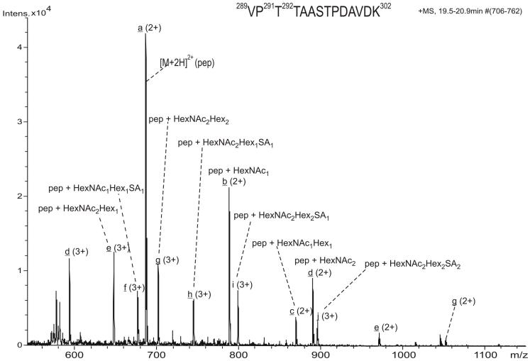
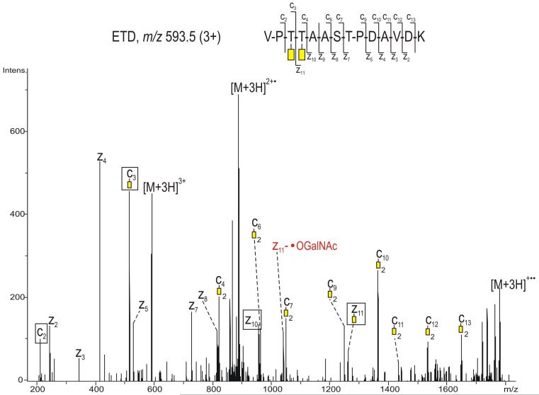
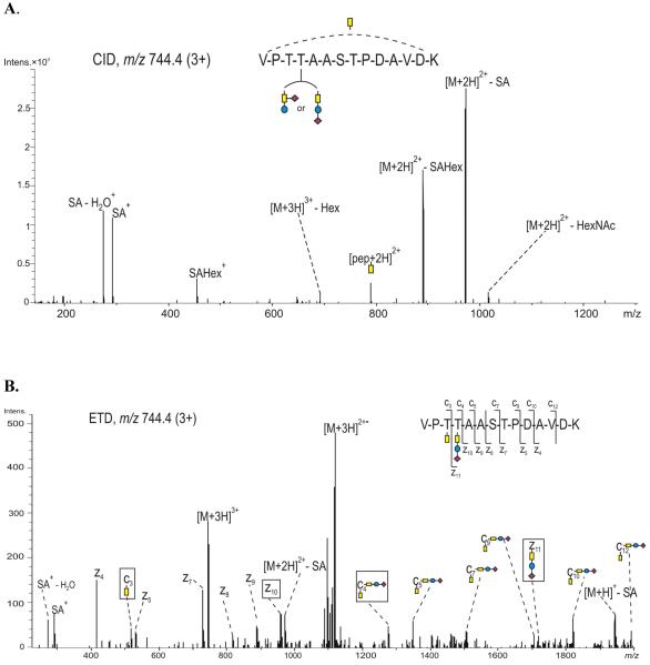
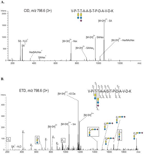
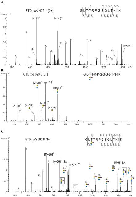
Accumulation and deposition of beta-amyloid peptide, a major constituent in neuritic plaques are hallmarks of Alzheimer's disease (AD) and AD-related neurodegenerative diseases. beta-Amyloid (Abeta) is derived from the proteolytic cleavage of amyloid precursor protein (APP), a transmembrane protein present in three major isoforms in brain comprising 695, 751 and 770 amino acids, respectively. Among other post-translational modifications, APP is modified during maturation by N- and O-glycosylation, which are thought to be responsible for its expression and secretion. Unlike N-glycosylation, no sites of O-glycosylation of APP have previously been reported. We report here the identification of three specific O-glycosylation sites of the secreted APP695 (sAPP695) produced in CHO cells, using a combination of high-performance liquid chromatography and electrospray-tandem mass spectrometry. With the use of electron transfer dissociation and collision induced dissociation (ETD and CID), we identified type, composition and structures of the Core 1 type O-linked glycans attached at the residues Thr 291, Thr 292 and Thr 576 of the full-length APP695. The glycosylations comprise multiple short glycans, containing N-acetyl galactosamine (GalNAc), Gal-GalNAc and sialic acid terminated structures. The presence of the glycopeptides in the tryptic mixture was identified using the CID-generated sugar oxonium ions. ETD proved to be valuable for the unambiguous identification of the modified sites as ETD fragmentation occurred along the peptide backbone with little or no cleavage of the glycans. Thus, the combination of the CID and ETD techniques in LC-MS is shown here, as a powerful tool for de novo identification of O-glycosylations at unknown modification sites in proteins.


Figures

Figure 1
Figure 1
Schematic representation of the non-amyloidogenic cleavage pathway of the transmembrane human APP695 generating the secreted fragment sAPPα and the C-terminal fragment CTFα; the amino acid sequence of sAPPα, residues 18 - 612, is depicted at the bottom. The N-glycosylation sites are highlighted in grey. The identified O-glycosylation sites are indicated in purple. The peptides derived from the use of different enzymes observed by LC-MS/MS are underlined.
Figure 2
Figure 2
Positive ion scan ESI mass spectrum summed over the chromatographic retention time 19.5 - 20.9 min, indicating the major glycoforms of the peptide 289-302 of the full length APP695. The individual glycopeptides are highlighted with letters from a through j. The charge states and the composition of the glycans determined for every glycopeptide are indicated for each glycoform.
Figure 3
Figure 3
(A.) ETD of the precursor ion m/z 788.2 (2+), corresponding to the peptide 289-302 of the full length APP695 containing the monosaccharide N-acetyl galactosamine (yellow rectangle) attached at Thr 292. The red label indicates the radical loss of 219Da (·OGalNAc) from the radical species [M+2H] and z11·, respectively. The fragment ions relevant for determination of the glycosylation site are indicated with black boxes. The inset in this spectrum shows the peak distribution of the species z7, z10 and z12 (see discussion in text). The spectrum was obtained using data dependent acquisition without supplemental ion activation. (B) Inset over the mass range m/z 1510 - 1580, showing the small neutral losses derived from the molecule radical ion of m/z 1576.4.
Figure 4
Figure 4
ETD of the precursor ion m/z 676.8 (3+), glycopeptide 289-302 of the full length APP695, showing the Core 1 type trisaccharide attached at Thr 292. The spectrum was obtained performing targeted MS/MS of the ion m/z 676.8 (3+), without supplemental ion activation. Color code: yellow - N-acetyl galactosamine, blue - galactose, purple - sialic acid. The fragment ions relevant for determination of the glycosylation site are indicated with black boxes.
Figure 5
Figure 5
ETD spectrum of the precursor ion m/z 593.5 (3+), corresponding to glycopeptide 289-302 of the full length APP695, showing each of the amino acids Thr 291 and Thr 292 occupied with N-acetyl galactosamine (yellow rectangle). The radical loss of 219 Da (·OGalNAc) from the z11 ion is indicated in red. The spectrum was obtained using data dependent acquisition without supplemental ion activation. The fragment ions relevant for determination of the glycosylation site(s) are indicated with black boxes.
Figure 6
Figure 6
(A) CID, and (B) ETD spectrum, of the precursor ion m/z 744.4 (3+), corresponding to glycopeptide 289-302 of the full length APP695, showing each of the amino acids Thr 291 and Thr 292 occupied with N-acetyl galactosamine and a Core 1 type trisaccharide, respectively. The spectrum was obtained using data dependent acquisition with a supplemental ion activation of 0.07 V. Color code: yellow - N-acetyl galactosamine, blue - galactose and purple - sialic acid. The fragment ions relevant for determination of the glycosylation site(s) are indicated with black boxes.
Figure 7
Figure 7
(A) CID and (B) ETD spectra of the precursor ion m/z 798.6 (3+) corresponding to glycopeptide 289-302 of the full length APP695, showing the amino acids Thr 291 and Thr 292 occupied with two distinct Core 1 type glycans. The spectrum was obtained using data dependent acquisition and the activation energy was 0.07 V. Color code: yellow - N-acetyl galactosamine, blue - galactose, purple - sialic acid. The fragment ions relevant for determination of the glycosylation site(s) are indicated with black boxes.
Figure 8
Figure 8
(A) ETD spectrum of the precursor ion m/z 472.1 (3+), showing the non-modified peptide 574-587 of the full length APP695 containing four potential O-glycosylation sites. (B) CID and (C) ETD of the precursor ion m/z 690.8 (3+), corresponding to glycopeptide 574-587 of the full length APP695, showing amino acid Thr 576 occupied with the indicated Core 1 type trisaccharide. The ETD spectrum was obtained using data dependent acquisition and the activation energy was 0.10 V. Color code: yellow - N-acetyl galactosamine, blue - galactose, purple - sialic acid. The fragment ions relevant for determination of the glycosylation site are indicated with black boxes.
Figure 9
Figure 9
Proposed structure types and linkages of identified O-glycosylations at Thr-291, Thr-292 and Thr-576.

References

    1. Gralle M, Ferreira ST. Structure and functions of the human amyloid precursor protein: the whole is more than the sum of its parts. Prog Neurobiol. 2007;82(1):11–32. - PubMed
    1. Goldgaber D, Lerman MI, McBride OW, Saffiotti U, Gajdusek DC. Characterization and chromosomal localization of a cDNA encoding brain amyloid of Alzheimer’s disease. Science. 1987;235(4791):877–80. - PubMed
    1. Palmert MR, Podlisny MB, Witker DS, Oltersdorf T, Younkin LH, Selkoe DJ, Younkin SG. The beta-amyloid protein precursor of Alzheimer disease has soluble derivatives found in human brain and cerebrospinal fluid. Proc Natl Acad Sci U S A. 1989;86(16):6338–42. - PMC - PubMed
    1. Lammich S, Kojro E, Postina R, Gilbert S, Pfeiffer R, Jasionowski M, Haass C, Fahrenholz F. Constitutive and regulated alpha-secretase cleavage of Alzheimer’s amyloid precursor protein by a disintegrin metalloprotease. Proc Natl Acad Sci U S A. 1999;96(7):3922–7. - PMC - PubMed
    1. Allinson TM, Parkin ET, Turner AJ, Hooper NM. ADAMs family members as amyloid precursor protein alpha-secretases. J Neurosci Res. 2003;74(3):342–52. - PubMed

Publication types

MeSH terms

Substances

LinkOut - more resources