

Review
doi: 10.1111/j.1365-2796.2008.02042.x.
Mucosal T-cell responses to HIV: responding at the front lines
Affiliations
- PMID: 19093960
- PMCID: PMC2827148
- DOI: 10.1111/j.1365-2796.2008.02042.x
Item in Clipboard
Review
Mucosal T-cell responses to HIV: responding at the front lines
J Intern Med.
2009 Jan.
Abstract
Mucosal surfaces of the body serve as the major portal of entry for human immunodeficiency virus (HIV). These tissues also house a majority of the body's lymphocytes, including the CD4(+) T cells that are the major cellular target for HIV infection. Mucosal surfaces are defended by innate and adaptive immune mechanisms, including secreted antibodies and CD8(+) cytotoxic T cells (CTL). CTL in mucosal lymphoid tissues may serve to limit viral replication, decreasing the host's viral burden as well as reducing the likelihood of sexual transmission to a naïve host. This review summarizes recent literature on HIV-specific T-cell responses in mucosal tissues, with an emphasis on the gastrointestinal tract.
Figures
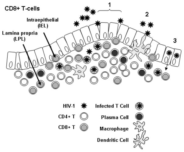
The figure shows an idealized rectal mucosa, with a single layer of columnar epithelium. HIV-1 may enter the mucosa through breaches in the epithelial layer (1); by binding to dendritic cell processes that extend through the epithelial layer to the lumen (2); via transcytosis across epithelial cells (3); or through a combination of mechanisms. CD8+ T-cells are present as intraepithelial cells (IEL) located between epithelial cells, and in the underlying layer as lamina propria lymphocytes (LPL). Also present in the lamina propria are CD4+ T-cells, macrophages, and antibody-producing plasma cells.
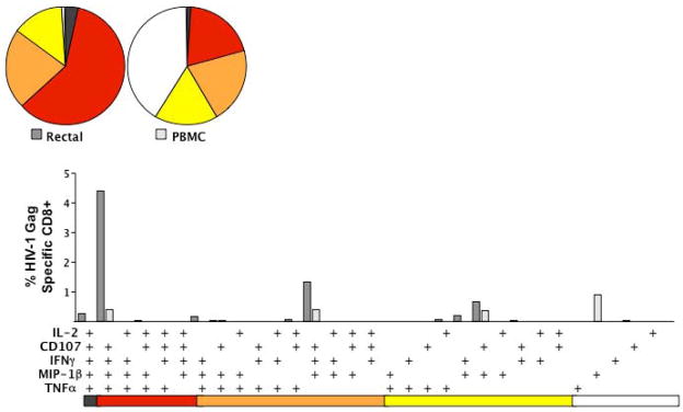
The figure summarizes data for one patient, an HIV controller whose Gag-specific CD8+ T-cell response is substantially stronger (in terms of percent responding cells) and more polyfunctional in rectal mucosa than in blood. Bar graphs show the magnitude of the Gag-specific response in each of 31 functional categories. The legend shows the combination of functions evaluated in each category. These combinations are color-coded based on the number of functions and correspond to wedges in the pie charts (5-function responses are black, 4-function red, 3-function orange, 2-function yellow, and single-function white). The rectal Gag-specific CD8+ T-cell response (white bars) of this HIV controller was heavily biased towards polyfunctional cells secreting CD107, MIP-1β, TNF-α and IFN-γ. In contrast, the Gag-specific response in PBMC was largely monofunctional. Data for over 20 HIV controllers, compared to non-controllers and patients on HAART, are reported elsewhere [62, 63].
Similar articles
-
Mucosal immunity to HIV: a review of recent literature.Curr Opin HIV AIDS. 2008 Sep;3(5):541-7. doi: 10.1097/COH.0b013e32830ab9ee. Curr Opin HIV AIDS. 2008. PMID: 19373019 Free PMC article.
-
Immune responses to HIV and SIV in mucosal tissues: 'location, location, location'.Curr Opin HIV AIDS. 2010 Mar;5(2):128-34. doi: 10.1097/COH.0b013e328335c178. Curr Opin HIV AIDS. 2010. PMID: 20543589 Free PMC article. Review.
-
The immunologic aspects of human immunodeficiency virus infection in the gastrointestinal tract.Semin Gastrointest Dis. 1996 Jan;7(1):19-29. Semin Gastrointest Dis. 1996. PMID: 8903576 Review.
-
Mucosal immunity in acute HIV: a review of recent work.Curr Opin HIV AIDS. 2025 May 1;20(3):193-198. doi: 10.1097/COH.0000000000000917. Epub 2025 Jan 24. Curr Opin HIV AIDS. 2025. PMID: 39903645 Review.
-
Viral suppression and immune restoration in the gastrointestinal mucosa of human immunodeficiency virus type 1-infected patients initiating therapy during primary or chronic infection.J Virol. 2006 Aug;80(16):8236-47. doi: 10.1128/JVI.00120-06. J Virol. 2006. PMID: 16873279 Free PMC article.
Cited by
-
Intraepithelial γδ T cells remain increased in the duodenum of AIDS patients despite antiretroviral treatment.PLoS One. 2012;7(1):e29066. doi: 10.1371/journal.pone.0029066. Epub 2012 Jan 4. PLoS One. 2012. PMID: 22238587 Free PMC article.
-
Camu Camu effects on microbial translocation and systemic immune activation in ART-treated people living with HIV: protocol of the single-arm non-randomised Camu Camu prebiotic pilot study (CIHR/CTN PT032).BMJ Open. 2022 Jan 17;12(1):e053081. doi: 10.1136/bmjopen-2021-053081. BMJ Open. 2022. PMID: 35039291 Free PMC article. Clinical Trial.
-
Pathogenic infection of Macaca nemestrina with a CCR5-tropic subtype-C simian-human immunodeficiency virus.Retrovirology. 2009 Jul 14;6:65. doi: 10.1186/1742-4690-6-65. Retrovirology. 2009. PMID: 19602283 Free PMC article.
-
Effect of metformin on the size of the HIV reservoir in non-diabetic ART-treated individuals: single-arm non-randomised Lilac pilot study protocol.BMJ Open. 2019 Apr 20;9(4):e028444. doi: 10.1136/bmjopen-2018-028444. BMJ Open. 2019. PMID: 31005944 Free PMC article.
-
Characterization of an effective CTL response against HIV and SIV infections.J Biomed Biotechnol. 2011;2011:103924. doi: 10.1155/2011/103924. Epub 2011 Sep 29. J Biomed Biotechnol. 2011. PMID: 21976964 Free PMC article. Review.
References
-
- Mowat A, Viney J. The anatomical basis of intestinal immunity. Immunol Rev. 1997;156:145–66. - PubMed
-
- Sansonetti PJ. War and peace at mucosal surfaces. Nat Rev Immunol. 2004;4:953–64. - PubMed
-
- Brandtzaeg P, Pabst R. Let’s go mucosal: communication on slippery ground. Trends Immunol. 2004;25:570–7. - PubMed
-
- Mowat AM. Anatomical basis of tolerance and immunity to intestinal antigens. Nat Rev Immunol. 2003;3:331–41. - PubMed
-
- Masopust D, Vezys V, Wherry EJ, Barber DL, Ahmed R. Cutting edge: gut microenvironment promotes differentiation of a unique memory CD8 T cell population. J Immunol. 2006;176:2079–83. - PubMed
Publication types
MeSH terms
Grants and funding
LinkOut - more resources
Full Text Sources
Medical
Research Materials
