Changes in prodynorphin gene expression and neuronal morphology in the hypothalamus of postmenopausal women
- PMID: 19094085
- PMCID: PMC2893873
- DOI: 10.1111/j.1365-2826.2008.01796.x
Changes in prodynorphin gene expression and neuronal morphology in the hypothalamus of postmenopausal women
Abstract
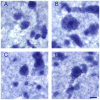
Human menopause is characterised by ovarian failure, gonadotrophin hypersecretion and hypertrophy of neurones expressing neurokinin B (NKB), kisspeptin (KiSS)-1 and oestrogen receptor (ER) alpha gene transcripts within the hypothalamic infundibular (arcuate) nucleus. In the arcuate nucleus of experimental animals, dynorphin, an opioid peptide, is colocalised with NKB, kisspeptin, ER alpha and progesterone receptors. Moreover, ovariectomy decreases the expression of prodynorphin gene transcripts in the arcuate nucleus of the ewe. Therefore, we hypothesised that the hypertrophied neurones in the infundibular nucleus of postmenopausal women would express prodynorphin mRNA and that menopause would be accompanied by changes in prodynorphin gene transcripts. In the present study, in situ hybridisation was performed on hypothalamic sections from premenopausal and postmenopausal women using a radiolabelled cDNA probe targeted to prodynorphin mRNA. Autoradiography and computer-assisted microscopy were used to map and count labelled neurones, measure neurone size and compare prodynorphin gene expression between premenopausal and postmenopausal groups. Neurones expressing dynorphin mRNA in the infundibular nucleus of the postmenopausal women were larger and exhibited hypertrophied morphological features. Moreover, there were fewer neurones labelled with the prodynorphin probe in the infundibular nucleus of the postmenopausal group compared to the premenopausal group. The number of dynorphin mRNA-expressing neurones was also reduced in the medial preoptic/anterior hypothalamic area of postmenopausal women without changes in cell size. No differences in cell number or size of dynorphin mRNA-expressing neurones were observed in any other hypothalamic region. Previous studies using animal models provide strong evidence that the changes in prodynorphin neuronal size and gene expression in postmenopausal women are secondary to the ovarian failure of menopause. Given the inhibitory effect of dynorphin on the reproductive axis, decreased dynorphin gene expression could play a role in the elevation in luteinising hormone secretion that occurs in postmenopausal women.
Figures


References
-
- Block E. Quantitative morphological investigations of the follicular system in women; variations at different ages. Acta Anat. 1952;14:108–123. - PubMed
-
- Hansen KR, Knowlton NS, Thyer AC, Charleston JS, Soules MR, Klein NA. A new model of reproductive aging: the decline in ovarian non-growing follicle number from birth to menopause. Hum Reprod. 2008;23:699–708. - PubMed
-
- Rance NE, McMullen NT, Smialek JE, Price DL, Young WS., III Postmenopausal hypertrophy of neurons expressing the estrogen receptor gene in the human hypothalamus. J Clin Endocrinol Metab. 1990;71:79–85. - PubMed
-
- Rance NE, Young WS., III Hypertrophy and increased gene expression of neurons containing neurokinin-B and substance-P messenger ribonucleic acids in the hypothalami of postmenopausal women. Endocrinology. 1991;128:2239–2247. - PubMed
Publication types
MeSH terms
Substances
Grants and funding
LinkOut - more resources
Full Text Sources
Other Literature Sources

