Comparative Study
. 2008 Dec;213(6):633-45.
doi: 10.1111/j.1469-7580.2008.01007.x.

Loss of translation elongation factor (eEF1A2) expression in vivo differentiates between Wallerian degeneration and dying-back neuronal pathology

Affiliations
Comparative Study

Loss of translation elongation factor (eEF1A2) expression in vivo differentiates between Wallerian degeneration and dying-back neuronal pathology

Lyndsay M Murray et al. J Anat. 2008 Dec.

Abstract

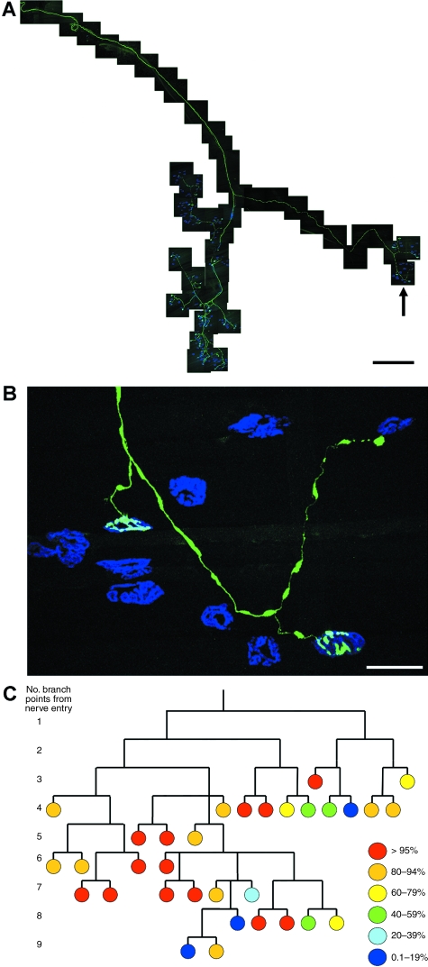
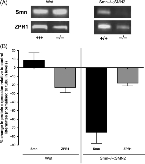
Wallerian degeneration and dying-back pathology are two well-known cellular pathways capable of regulating the breakdown and loss of axonal and synaptic compartments of neurons in vivo. However, the underlying mechanisms and molecular triggers of these pathways remain elusive. Here, we show that loss of translation elongation factor eEF1A2 expression in lower motor neurons and skeletal muscle fibres in homozygous Wasted mice triggered a dying-back neuropathy. Synaptic loss at the neuromuscular junction occurred in advance of axonal pathology and by a mechanism morphologically distinct from Wallerian degeneration. Dying-back pathology in Wasted mice was accompanied by reduced expression levels of the zinc finger protein ZPR1, as found in other dying-back neuropathies such as spinal muscular atrophy. Surprisingly, experimental nerve lesion revealed that Wallerian degeneration was significantly delayed in homozygous Wasted mice; morphological assessment revealed that approximately 80% of neuromuscular junctions in deep lumbrical muscles at 24 h and approximately 50% at 48 h had retained motor nerve terminals following tibial nerve lesion. This was in contrast to wild-type and heterozygous Wasted mice where < 5% of neuromuscular junctions had retained motor nerve terminals at 24 h post-lesion. These data show that eEF1A2 expression is required to prevent the initiation of dying-back pathology at the neuromuscular junction in vivo. In contrast, loss of eEF1A2 expression significantly inhibited the initiation and progression of Wallerian degeneration in vivo. We conclude that loss of eEF1A2 expression distinguishes mechanisms underlying dying-back pathology from those responsible for Wallerian degeneration in vivo and suggest that eEF1A2-dependent cascades may provide novel molecular targets to manipulate neurodegenerative pathways in lower motor neurons.


Figures

Fig. 1
Fig. 1
Synaptic loss at the NMJ in Wst mice occurs by a pathway morphologically distinct from WD. (A) Confocal micrograph showing a normal, control NMJ in an immunohistochemically-labelled transversus abdominis (TVA) muscle preparation from a P21 wild-type littermate mouse [green, 165 kDa neurofilaments and SV2; red, post-synaptic acetylcholine receptors labelled with α-bungarotoxin conjugated to tetramethylrhodamine isothiocyanate (TRITC-α-bungarotoxin)]. (B) Electron micrograph showing a normal, control NMJ in a lumbrical muscle preparation from a P25 wild-type littermate mouse. (C) Confocal micrograph showing two partially occupied NMJs in an immunohistochemically-labelled TVA muscle preparation from a P21 Wst mouse (green, 165 kDa neurofilaments and SV2; red, post-synaptic acetylcholine receptors labelled with TRITC-α-bungarotoxin). Partial occupancy of endplates is a morphological characteristic associated with nerve terminal retraction (e.g. dying-back) processes rather than WD. Electron micrographs showing partially occupied NMJs (identified by regions of vacant post-synaptic folds; blue arrows) in the flexor digitorum brevis (D) and lumbrical (E) muscles from late-symptomatic (P25) Wst mice. Note how the remaining motor nerve terminal boutons show none of the characteristic ultrastructural signs of WD; mitochondria, synaptic vesicles and pre-synaptic plasma membranes were all present and intact, and there was no evidence of phagocytosis by the terminal Schwann cell. (F) Confocal micrograph showing characteristic neurofilament accumulation, alongside interspersed thinning, in distal axons (blue and yellow arrows respectively) and motor nerve terminals from a P25 Wst mouse lumbrical muscle. (G) Electron micrograph showing a cross-sectional profile of an axon from the intramuscular region of an intercostal nerve supplying the TVA muscle in a P26 Wstmouse. Note the presence of large neurofilament accumulations in the axonal cytoplasm (cf. F) and the absence of any degenerative characteristics; the myelin sheath and axonal membranes remain intact. (H) Electron micrograph showing a NMJ in the flexor digitorum brevis muscle from a P26 Wstmouse. Note the presence of large neurofilament accumulations in the motor nerve terminal (blue arrows) but the lack of any morphological characteristics of WD (see above). (I) High-power electron micrograph showing three mitochondria in a motor nerve terminal from the flexor digitorum brevis muscle of a P26 Wstmouse. Note how the mitochondrial membranes and cristae are preserved (disruption of mitochondrial morphology is one of the earliest signs of WD). (J) Bar chart (mean ± SEM) showing significant increases in synaptic vesicle densities in motor nerve terminals from the lumbrical muscles of Wst mice at P20 (early-symptomatic) and P26 (late-symptomatic) compared with wild-type littermates (**P < 0.01 for both ages, anovawith Tukey's post-hoc test; N = 24 nerve terminals and n = 6694 vesicles in P20 wild-type, N = 20 and n = 3540 in P20 Wst, N = 26 and n = 8071 in P26 Wst). Synaptic vesicles are normally depleted from motor nerve terminals during WD. Scale bars: 10 µm (A), 15 µm (C), 0.75 µm (B, D and E), 20 µm (F), 0.75 µm (G), 1 µm (H), 0.1 µm (I).
Fig. 2
Fig. 2
Synaptic pathology extends to the flexor digitorum brevis (FDB) and levator auris longus (LAL) muscles in symptomatic Wst mice. Confocal micrographs showing NMJs in immunohistochemically-labelled LAL (A) and FDB (B) muscle preparations from P25 Wst mice (green, 165 kDa neurofilaments and SV2; red, post-synaptic acetylcholine receptors labelled with α-bungarotoxin conjugated to tetramethylrhodamine isothiocyanate). Partially occupied (white arrows in B) and vacant (white arrow in A) endplates were present throughout both muscle groups, as previously observed in transversus abdominis (TVA) and lumbrical muscles (Fig. 1 in the current paper; Newbery et al. 2005). Occasionally, remnants of synaptic terminals and distal axons withdrawing from post-synaptic endplates were observed (‘retraction bulbs’; blue arrow in A). (C) Bar chart (mean ± SEM) showing the percentage of fully occupied endplates, partially occupied endplates and vacant endplates in LAL muscles from wild-type mice at P20 (WT P20; black bars; N = 2 mice), Wst mice at P20 (−/− P20; N = 3 mice) and Wst mice at P25 (−/− P25; N = 4 mice). Note how the percentage of fully occupied endplates declines and the percentage of vacant endplates increases with advancing age in Wst mice. Similar loss of pre-synaptic innervation was observed in FDB muscles at P25, albeit to a lesser extent than in LAL or TVA (as in neighbouring lumbrical muscles; ∼30–40% of NMJs showed disrupted morphology; data not shown). Scale bars: 20 µm (A), 15 µm (B).
Fig. 3
Fig. 3
Functional loss precedes structural loss at NMJs undergoing dying-back pathology in Wst mice. Confocal micrograph (merge of all three channels in top panel with separate channels shown below) of NMJs from a P25 Wst;YFP-H mouse LAL muscle. Motor nerve terminals were loaded with FM4–64FX using a depolarizing high-potassium solution leading to the selective labelling of functionally-active terminals (i.e. with the retained ability to recycle synaptic vesicles). In the preparation shown, two NMJs with motor nerve terminals containing YFP can be seen but only one of these motor nerve terminals demonstrated retained functional ability (red arrow; note the presence of strong FM4–64FX label in the bottom panel). The junction indicated by the white arrow was present morphologically (as shown by the YFP label) but had lost its functional capacity to recycle synaptic vesicles and hence had not taken up the FM4–64FX label. Thus, synaptic function was lost before morphological retraction occurred in Wst mice. By contrast, in control preparations from YFP-H wild-type littermates, all motor nerve terminals showed strong FM4–64FX labelling (data not shown). Scale bar, 35 µm.
Fig. 4
Fig. 4
Synaptic pathology occurs in the absence of axonal pathology in symptomatic Wst mice. Montage of fluorescence micrographs showing YFP-labelled axons in an intercostal nerve (A) (supplying the transversus abdominis muscle) and a tibial nerve (B) (supplying the lumbrical muscles) from P25 Wst;YFP-H mice. Note that not all axons are labelled with YFP, allowing direct visualization and tracing of individual axons for several hundred microns. All labelled axons were intact over the entire length reconstructed in both nerves. This is in stark contrast to the fragmented appearance of axons undergoing WD (cf. Beirowski et al. 2005). (C) High-power fluorescence micrograph of an individual YFP-labelled axon in an intercostal nerve from a P25 Wst;YFP-H mouse showing intact axonal morphology. (D) Cross-section of a toluidine blue-stained intercostal nerve from a P24 Wst mouse. Less than 1% of axons examined using this approach showed any morphological signs of degeneration. (E) Electron micrograph of axons from an intramuscular region of intercostal nerve from a P26 Wst mouse, supporting analysis of toluidine blue-stained sections (D) by showing no evidence of axonal degeneration. All axons examined had normal myelin sheaths and axonal membranes. Scale bars: 200 µm (A), 450 µm (B), 25 µm (C), 35 µm (D), 4 µm (E).
Fig. 5
Fig. 5
Asynchronous loss of synapses at the NMJ within single motor units in late-symptomatic Wst mice. (A) Montage of confocal micrographs showing a single YFP-H-labelled motor unit from a P25 Wst;YFP-H mouse (green, YFP; blue, post-synaptic acetylcholine receptors labelled with α-bungarotoxin). (B) Higher power confocal micrograph showing a small region of the larger whole motor unit shown in A (position indicated by black arrow in A). Note how the first (far left) NMJ is fully occupied, the second (lower right) NMJ is partially occupied and the third (upper right) NMJ has lost its pre-synaptic input, remnants of which are left as a retraction bulb. (C) Branch diagram generated from an analysis of the single motor unit shown in A, with each individual NMJ represented as a circle relative to its branch point position within the motor unit. The occupancy status of each NMJ is represented by the colour of the circle (key to colours is on the right of the panel). Note how synaptic retraction occurred asynchronously throughout the motor unit, with a full spread of occupancies present. Note also how fully occupied (i.e. ‘normal’; red) NMJs were distributed at locations throughout the motor unit, as were almost fully retracted NMJs (blue). Scale bars: 500 µm (A), 50 µm (B).
Fig. 6
Fig. 6
Dying-back pathology in Wst mice occurs independently of changes in Smn protein expression but correlates with modest reductions in ZPR1 protein levels. (A) Representative fluorescent western blots showing levels of Smn and ZPR1 protein in Wst mice vs. control littermates (left panel) and Smn–/–;SMN2 mice vs. control littermates (right panel). (B) Bar chart (mean ± SEM) showing expression levels of Smn and ZPR1 protein in the mid-thoracic spinal cord of late-symptomatic Wst (left bars; P25) and Smn–/–;SMN2 (right bars; P5) mice compared with respective control littermate mice, quantified using fluorescent western blots (N = 3 mice per genotype). Smn protein levels did not decrease in Wst mice (if anything showing a modest increase in expression) but, as expected, decreased by around 75% in Smn–/–;SMN2 mice. Thus, the dying-back pathology observed in Wst mice is not simply occurring due to reduced levels of Smn protein. ZPR1 protein levels, however, were reduced by similar amounts in both Wst and Smn–/–;SMN2 mice.
Fig. 7
Fig. 7
eEF1A2 expression is required for the normal initiation and progression of axotomy-induced WD. Confocal micrographs showing NMJs in immunohistochemically-labelled lumbrical muscle preparations from P25 wild-type (+/+) (A) and homozygous Wst (−/−) (B) mice at 24 h after a tibial nerve cut (green, 150 kDa neurofilaments; red, post-synaptic acetylcholine receptors). As expected, axotomy resulted in almost complete WD of motor nerve terminals and distal axon collaterals in lumbrical muscles of wild-type mice (A). Surprisingly, however, axotomy-induced WD was almost completely absent from homozygous Wst mice at the same time-point (B). (C) Montage of fluorescence micrographs showing YFP-labelled axons in a tibial nerve (supplying the lumbrical muscles) from a P25 Wst;YFP-H mouse at 24 h after a tibial nerve cut. Note the complete absence of any axonal pathology at this time-point. As expected, similar data were obtained from +/+ mice as gross morphological features of axonal WD do not begin to appear until 24 h after lesion (cf. Beirowski et al. 2005; data not shown). (D) Electron micrograph showing early ultrastructural signs of WD in +/+ tibial nerves at 24 h after axotomy (swollen and disrupted mitochondria; red arrows). (E) Electron micrograph showing lack of early ultrastructural signs of WD in −/− tibial nerves at 24 h after axotomy (mitochondria remained intact; red arrows). (F) Bar chart (mean ± SEM) showing the percentage of NMJs remaining in lumbrical muscles at 24 h post-axotomy (***P < 0.001; Kruskal Wallis test with Dunn's post-hoc; n = 57 muscles, N = 19 mice +/+; n = 36 muscles, N = 12 mice +/−; n = 36 muscles, N = 12 mice −/−). Note how < 5% of NMJs remained intact in muscles from wild-type mice, whereas ∼80% of NMJs remained intact in muscles from homozygous Wst mice. (G) Bar chart (mean ± SEM) showing the percentage of NMJs remaining in lumbrical muscles from homozygous Wst mice at 24 and 48 h post-axotomy (n = 36 muscles, N = 12 mice −/− 24 h; n = 6 muscles, N = 2 mice −/− 48 h). Note how ∼50% of NMJs remained intact in muscles from homozygous Wst mice even at 48 h post-axotomy. Scale bars: 50 µm (A and B), 200 µm (C), 2 µm (D and E).

Similar articles

Cited by

References

    1. Balice-Gordon RJ, Smith DB, Goldman J, et al. Functional motor unit failure precedes neuromuscular degeneration in canine motor neuron disease. Ann Neurol. 2000;47:596–605. - PubMed
    1. Baxter B, Gillingwater TH, Parson SH. Rapid loss of motor nerve terminals following hypoxia-reperfusion injury occurs via mechanisms distinct from classic Wallerian degeneration. J Anat. 2008;212:827–835. - PMC - PubMed
    1. Beirowski B, Adalbert R, Wagner D, et al. The progressive nature of Wallerian degeneration in wild-type and slow Wallerian degeneration (WldS) nerves. BMC Neurosci. 2005;6(1):6. - PMC - PubMed
    1. Bettini NL, Moores TS, Baxter B, Deuchars J, Parson SH. Dynamic remodelling of synapses can occur in the absence of the parent cell body. BMC Neurosci. 2007;8:79. - PMC - PubMed
    1. Chambers DM, Peters J, Abbott CM. The lethal mutation of the mouse wasted (wst) is a deletion that abolishes expression of a tissue-specific isoform of translation elongation factor 1alpha, encoded by the Eef1a2 gene. Proc Natl Acad Sci USA. 1998;95:4463–4468. - PMC - PubMed

Publication types

Substances

LinkOut - more resources