. 2008 Dec;213(6):718-24.
doi: 10.1111/j.1469-7580.2008.00997.x.

Development of the human Achilles tendon enthesis organ

Affiliations

Development of the human Achilles tendon enthesis organ

H M Shaw et al. J Anat. 2008 Dec.

Abstract

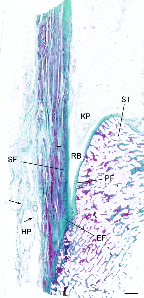
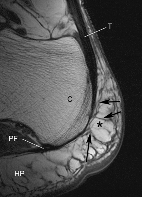
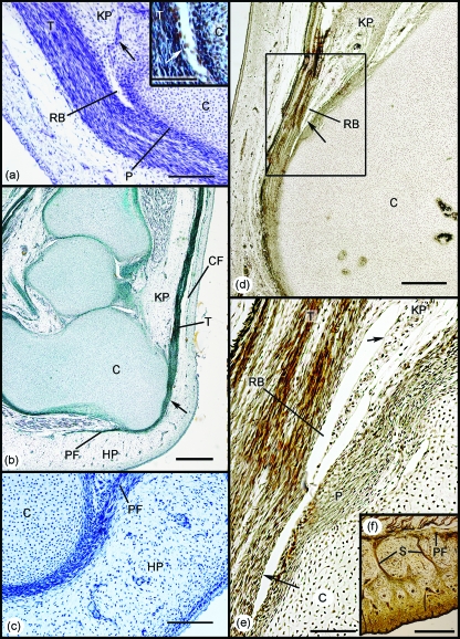
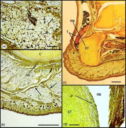
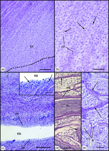
The attachment of the Achilles tendon is part of an 'enthesis organ' that reduces stress concentration at the hard-soft tissue interface. The organ also includes opposing sesamoid and periosteal fibrocartilages, a bursa and Kager's fat pad. In addition, the deep crural and plantar fasciae contribute to Achilles stress dissipation and could also be regarded as components. Here we describe the sequence in which these various tissues differentiate. Serial sections of feet from spontaneously aborted foetuses (crown rump lengths 22-322 mm) were examined. All slides formed part of an existing collection of histologically sectioned embryological material, obtained under Spanish law and housed in the Universidad Complutense, Madrid. From the earliest stages, it was evident that the Achilles tendon and plantar fascia had a mutual attachment to the calcaneal perichondrium. The first components of the enthesis organ to appear (in the 45-mm foetus) were the retrocalcaneal bursa and the crural fascia. The former developed by cavitation within the mesenchyme that later gave rise to Kager's fat pad. The tip of the putative fat pad protruded into the developing bursa in the 110-mm foetus and fully differentiated adipocytes were apparent in the 17-mm foetus. All three fibrocartilages were first recognisable in the 332-mm foetus--at which time adipogenesis had commenced in the heel fat pad. The sequence in which the various elements became apparent suggests that bursal formation and the appearance of the crural fascia may be necessary to facilitate the foot movements that subsequently lead to fibrocartilage differentiation. The later commencement of adipogenesis in the heel than in Kager's pad probably reflects the non-weight environment in utero. The direct continuity between plantar fascia and Achilles tendon that is characteristic of the adult reflects the initial attachment of both structures to the calcaneal perichondrium rather than to the skeletal anlagen itself.


Figures

Fig. 1
Fig. 1
The enthesis organ of the adult Achilles tendon. The enthesis itself is characterised by an enthesis fibrocartilage (EF) which is most prominent in the superior part of the attachment. In addition, there is a sesamoid fibrocartilage (SF) in the deep surface of the tendon, immediately adjacent to the enthesis, and a corresponding periosteal fibrocartilage (PF) on the opposing surface of the superior tuberosity (ST) of the calcaneus. The tendon (T) and bone are separated by the retrocalcaneal bursa (RB) into which protrudes the tip of Kager's fat pad (KP). Note that the heel fat pad (HP) extends upwards from the sole of the foot to the posterior part of the Achilles tendon. Arrows, fibrous septa of the fat pad. Masson's trichrome. Scale bar: 2 mm.
Fig. 2
Fig. 2
A non-fat suppressed sagittal MR image of the normal heel of a young female volunteer (age 42 years) showing how the Achilles tendon (T) is directly continuous with the plantar fascia (PF) around the posterior aspect of the calcaneus (C). Fat (asterisk) is high signal (bright). Tendons, fascia and septa are low signal (black). Note how the heel fat pad (HP) extends upwards behind the tendon and that its fibrous septa are connected to it (arrows).
Fig. 3
Fig. 3
Development of the enthesis organ in 45–110-mm foetuses. (a) Initially the Achilles tendon (T) attaches to the perichondrium (P) of the calcaneus (C). The earliest sign of the differentiation of the enthesis organ beyond the establishment of the enthesis itself, was the development of the retrocalcaneal bursa (RB) by cavitation within the vascular mesenchymal tissue occupying the position of the future Kager's fat pad (KP). Arrow – blood vessels. 45-mm foetus. Hansen's hematoxylin. Scale bar: 100 µm. Inset: Higher power view of the developing bursa to show the appearance of a layer of flattened mesenchymal cells (putative synovium – arrow). (b) The continuity of the Achilles tendon (T) and the plantar fascia (PF) is strikingly evident in this 57-mm foetus (arrow). A condensation of mesenchyme marks the first evidence of differentiation of the crural fascia (CF), but there is no adipogenesis in the region of the future Kager's (KP) or putative heel (HP) pads. Azan. Scale bar: 500 µm. (c) Higher power view of the region of the developing heel pad in a 45-mm foetus. Note that as in (b), the heel pad (HP) is purely mesenchymal and there is no evidence of the characteristic fibrous septa of the adult. (d) In the 110-mm foetus, the tip (arrow) of the future Kager's fat pad (KP) can now be seen to protrude into the retrocalcaneal bursa (RB). C, calcaneus. Bielchowsky. Scale bar: 500 µm. (e) Enlargement of the region in the rectangle in (d). The entire bursa (RB) is lined with a single layer of synovial cells (arrows). C, calcaneus; KP, Kager's fat pad; P, perichondrium; T, tendon. (f) The heel region of a 110-mm foetus. Prominent fibrous septa (S) are obvious, but adipogenesis has not commenced.
Fig. 4
Fig. 4
Development of the enthesis organ in the 177-mm foetus. (a) Adipocytes were first identifiable within Kager's fat pad at this stage (arrows). Scale bar:100 µm. (b) Although fat cells had not yet differentiated in the heel pad, fibrous septa were conspicuous (arrows). C, calcaneus; PF, plantar fascia. Scale bar: 500 µm. (c) A superior tuberosity (ST) had now developed on the posterior aspect of the calcaneus adjacent to the retrocalcaneal bursa (RB). Note also how the heel pad extends upwards behind the Achilles tendon (T) in the foetus as well as the adult (compare with Fig. 2). Scale bar: 2 mm. (d) Although the perichondrium (P) covering the superior tuberosity (ST) was greatly thickened, periosteal fibrocartilage had not yet differentiated. RB, retrocalcaneal bursa; T, tendon. Scale bar: 100 µm. All sections were stained with Bielchowsky's silver stain.
Fig. 5
Fig. 5
Development of the enthesis organ in the 332-mm foetus. (a) The enthesis fibrocartilage (EF) was first identifiable at this stage. Note the gradual bending of collagen fibres, as the Achilles tendon (T) inserts into the cartilaginous calcaneus (C). Scale bar: 100 µm. (b) Higher power view of the enthesis fibrocartilage in (a). Note the characteristic rounded fibrocartilage cells (arrows) interspersed between small bundles of collagen fibres (CF). Scale bar: 50 µm. (c) Both the sesamoid (SF) and periosteal (PF) fibrocartilages were also evident in the 332-mm foetus – forming the walls of the distal part of the retrocalcaneal bursa (RB). C, calcaneus. Scale bar: 100 µm. Inset: Higher power view of the sesamoid fibrocartilage in (c). Note the characteristic rounded cells (arrows). Scale bar: 100 µm. (d) Large myelinated nerve fascicles (N) surrounded by a prominent perineurium (PN) were evident within Kager's fat pad at this stage. Collections of well-differentiated adipocytes were separated by thick fibrous septa (arrows). Scale bar: 100 µm. (e) At this stage of development, adipogenesis was evident for the first time in the heel pad. Note the organisation of adipocytes (A) into locules, separated by thick fibrous septa (arrows). Scale bar: 100 µm. All slides were stained with Hansen's hematoxylin.

References

    1. Andersen H. Histochemistry and development of the human shoulder and acromioclavicular joints with particular reference to the early development of the clavicle. Acta Anat (Basel) 1963;55:124–165. - PubMed
    1. Andersen H, Bro-Rasmussen F. Histochemical studies on the histogenesis of the joints in human fetuses with special reference to the development of the joint cavities in the hand and foot. Am J Anat. 1961;108:111–122.
    1. Archer CW, Dowthwaite GP, Francis-West P. Development of synovial joints. Birth Defects Res C Embryo Today. 2003;69:144–155. - PubMed
    1. Benjamin M, McGonagle D. The anatomical basis for disease localisation in seronegative spondyloarthropathy at entheses and related sites. J Anat. 2001;199:503–526. - PMC - PubMed
    1. Benjamin M, McGonagle D. Histopathologic changes at ‘synovio–entheseal complexes’ suggesting a novel mechanism for synovitis in osteoarthritis and spondylarthritis. Arthritis Rheum. 2007;56:3601–3609. - PubMed

Publication types