. 2009 Mar 1;45(1):17-28.
doi: 10.1016/j.neuroimage.2008.11.010. Epub 2008 Nov 25.

Loss of cerebral white matter structural integrity tracks the gray matter metabolic decline in normal aging

Affiliations

Loss of cerebral white matter structural integrity tracks the gray matter metabolic decline in normal aging

P Kochunov et al. Neuroimage. .

Abstract

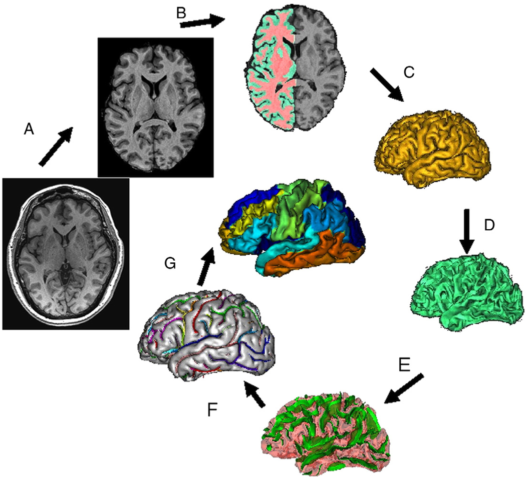
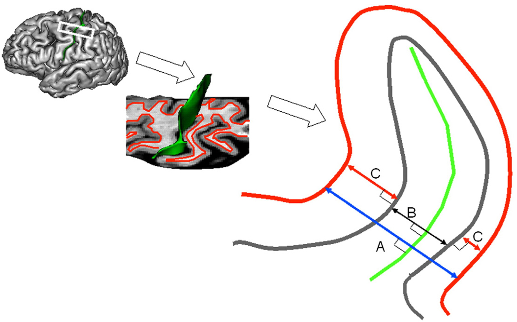
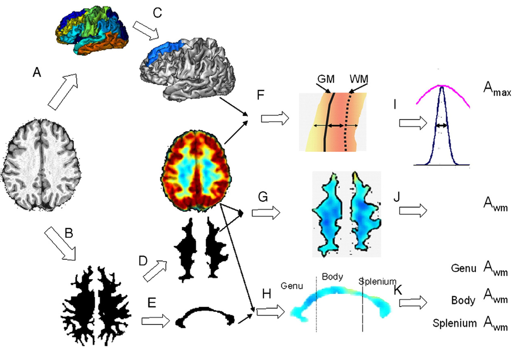
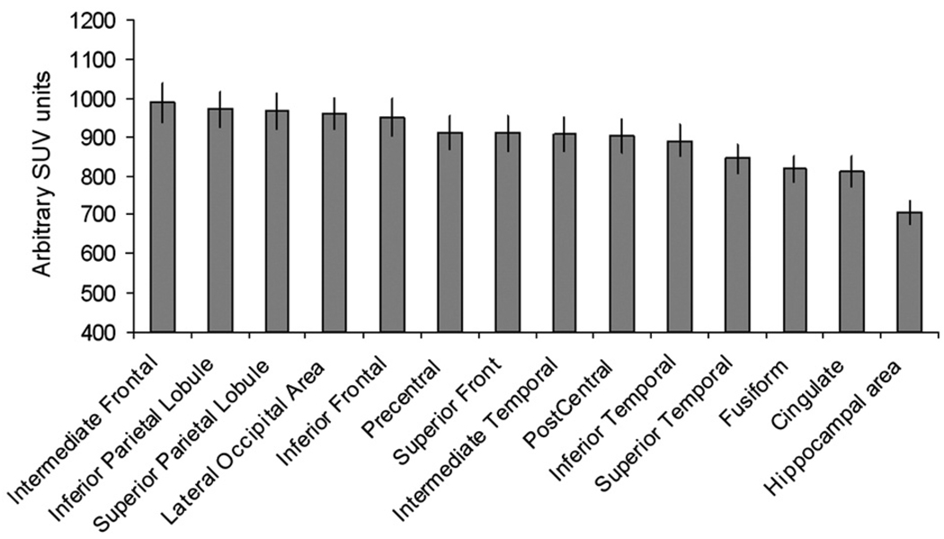
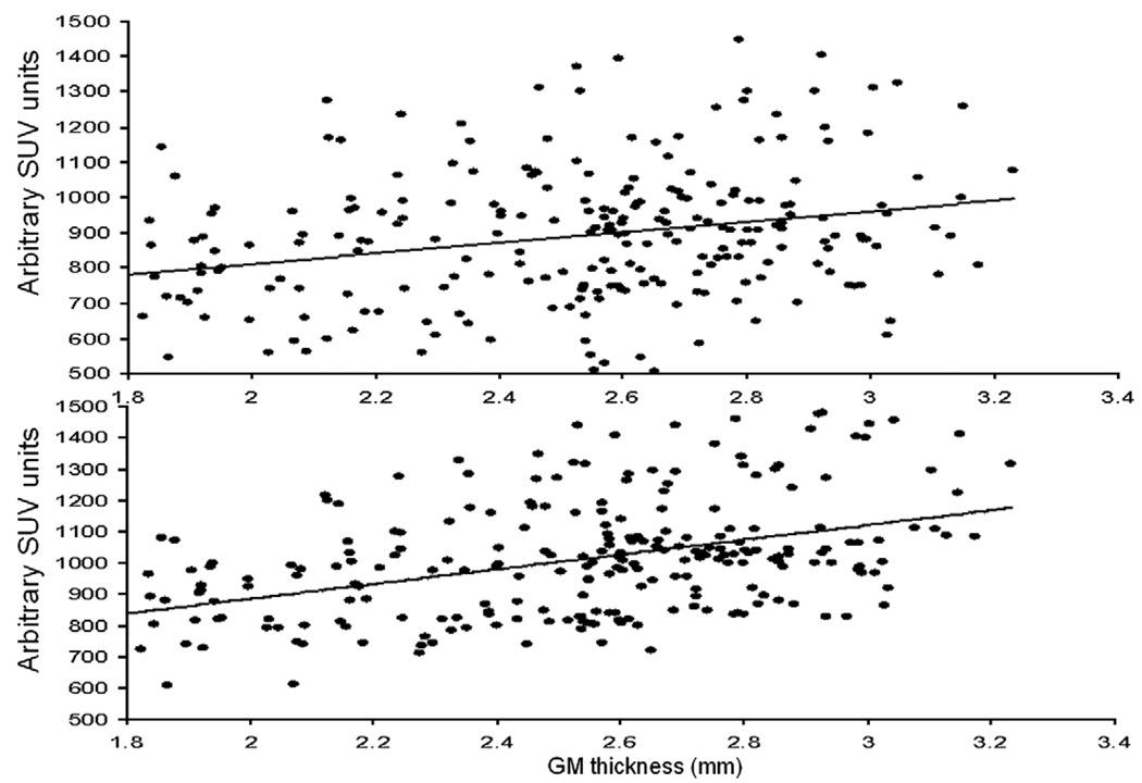
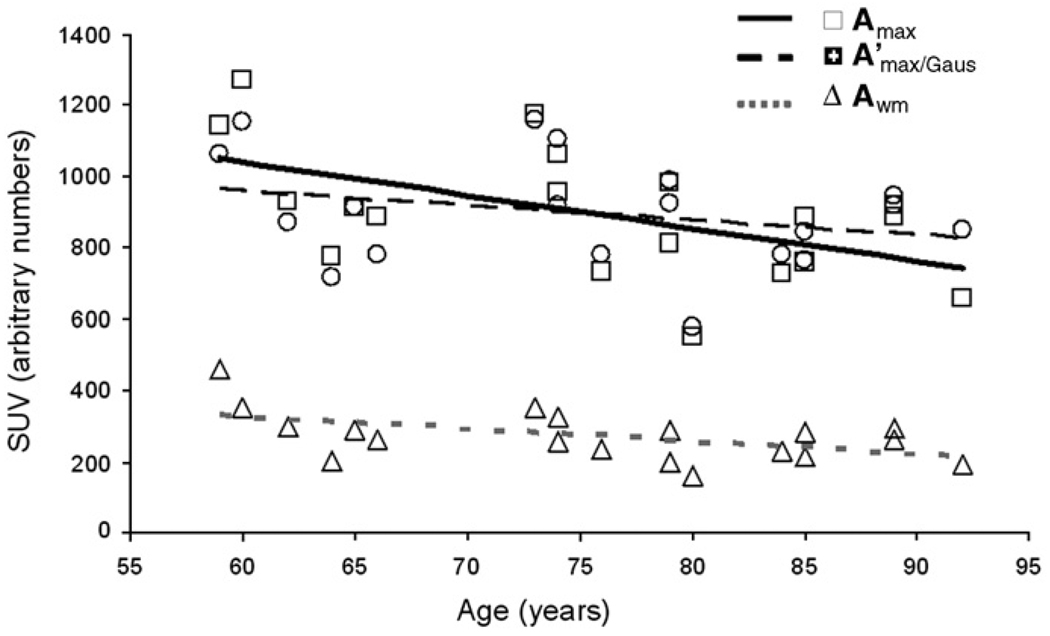
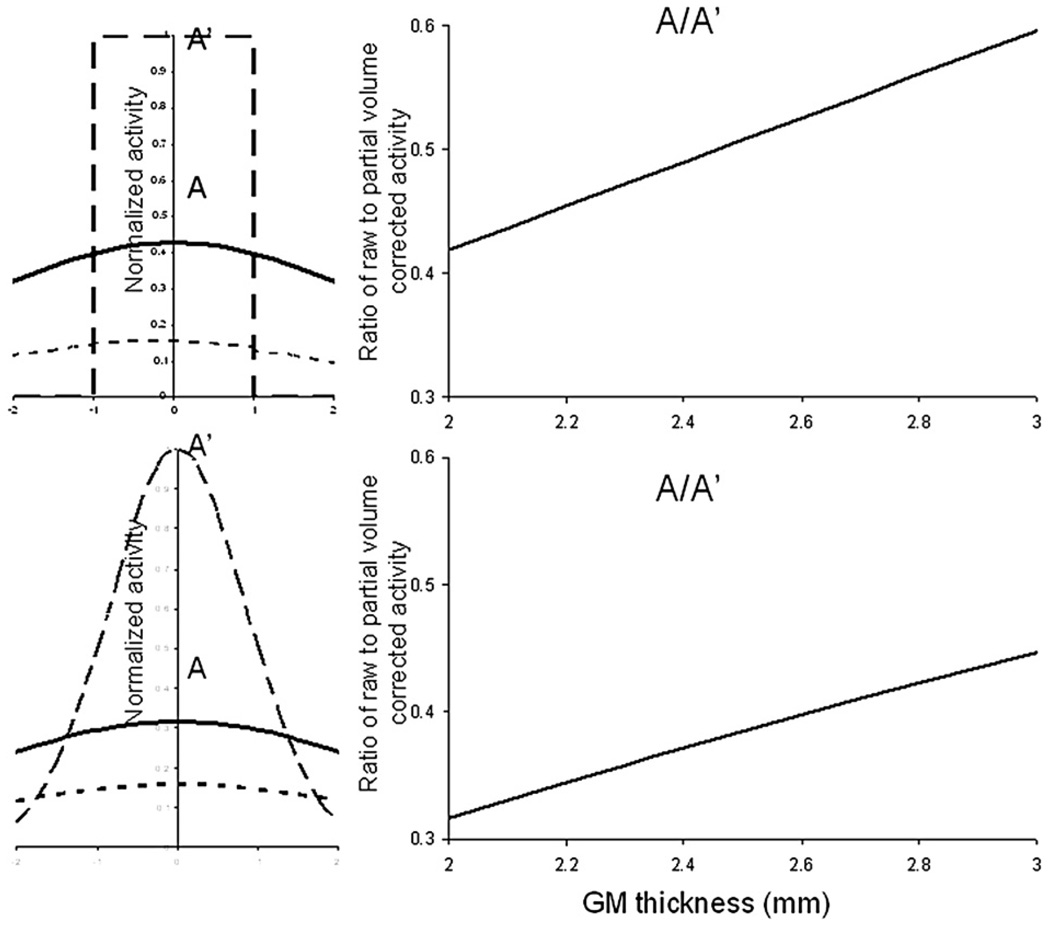
Relationships between structural MRI-based markers of declining cerebral integrity, and regional PET measurements of (18)FDG uptake have not been studied well in normal aging. In this manuscript we relate changes in cerebral morphology to regional cerebral glucose uptake for 14 major cortical areas in 19 healthy older individuals (age 59-92 years). Measurements of cerebral integrity included gray matter (GM) thickness, sulcal and intergyral spans, fractional anisotropy (FA) of water diffusion and volume of hyperintense WM (HWM) lesions. (18)FDG-PET measurements were converted to standard uptake values and corrected for partial volume artifact. Following this, cortical FDG uptake was significantly correlated with several indices of WM integrity that we previously observed to be sensitive to cognitive decline in executive function, including intergyral span and HWM volumes. Our findings suggest that the age-related decline in white matter integrity, observed as increases in HWM lesions, intergyral spans and reduction in FA, correlated with a decline in the global and regional cerebral glucose uptake. Our findings support the emerging consensus that WM integrity indices are sensitive predictors of declining cerebral health in normal aging. Specifically, age-related WM degradation in the thinly myelinated association tracts appears to track the decreases in global and regional rates of glucose uptake.


Figures

Fig. 1
Fig. 1
T1-w image processing pipelines. A T1-w image is skull-stripped, globally spatially normalized, and RF-inhomogeneity corrected (A). Next, cerebral hemispheres and cerebellum and identified and tissue classified (B); cortical surfaces for GM and WM are calculated (C, D) and homotopic erosion operation and crevasse detector are used to reconstruct sulcal surface as the medial surface of the two opposing gyral banks (E). Sulcal identification pipeline uses a congregation of 500 artificial neural network-based pattern classifiers to identify (F) sulcal landmarks and to perform gyral segmentation of the cortex (G).
Fig. 2
Fig. 2
Cortical markers of cerebral integrity. T1-w image processing pipeline (Fig. 1) produced sulcal surface (green) and pial GM (gray) and inner WM (red) surfaces. For each sulcus three average measurements of cortical cerebral integrity are computed: average intergyral (A), sulcal (B) spans and average GM thickness (C). Intergyral span (A) is defined as the 3-D distance between opposing points on the WM mesh along the normal projection to the sulcal surface. Sulcal span (B) is defined as a 3D distance between opposing points on the GM mesh along the normal projections to the sulcal surface. Gyral GM thickness (C) is defined as a distance between pial GM and inner WM meshes at the direction normal to the pial GM surface. These measurements are calculated for every vertex of the sulcal surface resulting in averaging of ~2–5000 measurements for each sulcus.
Fig. 3
Fig. 3
Extraction of regional FDG PET measurements. Anatomical images were processed with BV pipeline that produced cortical parcelation maps (A) and mask of cerebral WM (B). For each gyrus/cortical area (C), crossectional FDG intensity and WM/GM distance were analyzed to measure peak intensity Amax and GM thickness (F, I). These measurements were performed for each of the nodes of the gyrus (~10000) and results were averaged. The cerebral WM mask was eroded (D) to measure Awm, average WM activity. The mask of the corpus callosum (CC) was eroded (E) to measure Awm for genu, body and splenium of CC.
Fig. 4
Fig. 4
Baseline FDG uptake values (average Amax) for 14 cortical gyri/areas.
Fig. 5
Fig. 5
Cortical FDG uptake (Amax) values for 14 cortical gyri/areas plotted vs. GM thickness for 19 subjects before (top) and after intensity normalization (bottom) that normalized the average FDG intensity for every gyrus/area.
Fig. 6
Fig. 6
Whole-brain cerebral metabolism markers plotted vs. age. Regression lines are shown for uncorrected cortical GM (solid), corrected cortical GM (interrupted) and cerebral WM (shaded, dotted) average FDG uptake values.
Fig. S1
Fig. S1
Partial voxel averaging correction was modeled using rectangular (top) and Gaussian (bottom) approximations. The graphs are shown for FWHM of the PET camera set at 6 mm.

Similar articles

Cited by

References

    1. Abe O, Aoki S, Hayashi N, Yamada H, Kunimatsu A, Mori H, Yoshikawa T, Okubo T, Ohtomo K. Normal aging in the central nervous system: quantitative MR diffusion-tensor analysis. Neurobiol. Aging. 2002;23:433–441. - PubMed
    1. Aboitiz F. Brain connections: interhemispheric fiber systems and anatomical brain asymmetries in humans. Biol. Res. 1992;25:51–61. - PubMed
    1. Aboitiz F, Scheibel AB, Fisher RS, Zaidel E. Fiber composition of the human corpus callosum. Brain Res. 1992;598:143–153. - PubMed
    1. Bakshi R, Caruthers SD, Janardhan V, Wasay M. Intraventricular CSF pulsation artifact on fast fluid-attenuated inversion-recovery MR images: analysis of 100 consecutive normal studies. AJNR Am. J. Neuroradiol. 2000;21:503–508. - PMC - PubMed
    1. Bartzokis G. Age-related myelin breakdown: a developmental model of cognitive decline and Alzheimer's disease. Neurobiol. Aging. 2004;25:5–18. - PubMed

Publication types

Substances