. 2009 Feb 20;284(8):4881-8.
doi: 10.1074/jbc.M808077200. Epub 2008 Dec 18.

Regulation of C-type lectin antimicrobial activity by a flexible N-terminal prosegment

Affiliations

Regulation of C-type lectin antimicrobial activity by a flexible N-terminal prosegment

Sohini Mukherjee et al. J Biol Chem. .

Abstract

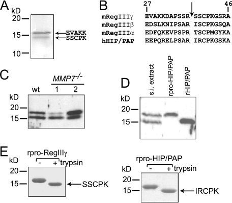
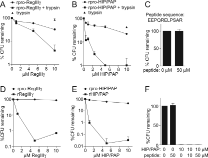
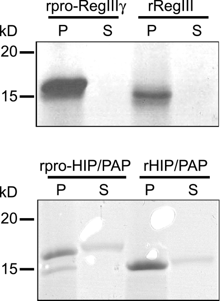
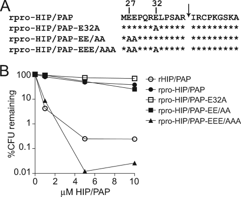
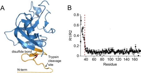
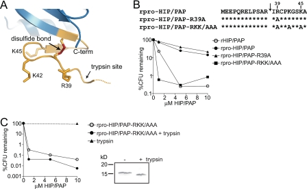
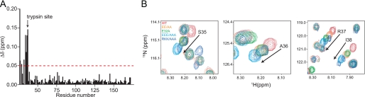
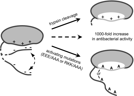
Members of the RegIII family of intestinal C-type lectins are directly antibacterial proteins that play a vital role in maintaining host-bacterial homeostasis in the mammalian gut, yet little is known about the mechanisms that regulate their biological activity. Here we show that the antibacterial activities of mouse RegIIIgamma and its human ortholog, HIP/PAP, are tightly controlled by an inhibitory N-terminal prosegment that is removed by trypsin in vivo. NMR spectroscopy revealed a high degree of conformational flexibility in the HIP/PAP inhibitory prosegment, and mutation of either acidic prosegment residues or basic core protein residues disrupted prosegment inhibitory activity. NMR analyses of pro-HIP/PAP variants revealed distinctive colinear backbone amide chemical shift changes that correlated with antibacterial activity, suggesting that prosegment-HIP/PAP interactions are linked to a two-state conformational switch between biologically active and inactive protein states. These findings reveal a novel regulatory mechanism governing C-type lectin biological function and yield new insight into the control of intestinal innate immunity.


Figures

FIGURE 1.
FIGURE 1.
RegIIIγ is proteolytically processed by trypsin in vivo. A, purification and N-terminal sequencing of endogenous mouse RegIIIγ reveals processing at the conserved N-terminal trypsin site. B, conserved canonical trypsin site (indicated by arrow) is present near the N terminus of mouse and human RegIII family members. Residue numbers are based on the deduced sequence which includes the signal peptide. Position 1 corresponds to the initiating methionine. C, MMP-7 is dispensable for RegIIIγ processing. 20 μg of protein extract from wild-type and MMP7–/– mice were immunoblotted with anti-RegIIIγ antibody. D, evidence for in vivo proteolytic processing of HIP/PAP. 20 μg of human intestinal protein extract was immunoblotted and probed with anti-RegIIIγ antiserum. Recombinant pro-HIP/PAP (rpro-HIP/PAP; with the N-terminal signal sequence replaced by methionine) and recombinant processed HIP/PAP (rHIP/PAP; with the N-terminal tryptic fragment replaced by methionine) were included for size comparison. s.i., small intestinal E, in vitro incubation of purified recombinant pro-RegIIIγ (rpro-RegIIIγ) and rpro-HIP/PAP with bovine trypsin results in quantitative cleavage at the conserved trypsin site to yield a homogeneous product. Proteins were digested with a 1:200 molar ratio of trypsin:lectin and were analyzed by SDS-PAGE. N-terminal sequencing verified cleavage at Arg37–Ser38 and Arg37–Ile38, respectively.
FIGURE 2.
FIGURE 2.
Proteolysis of the N terminus by trypsin activates lectin antibacterial activity. A and B, trypsin proteolysis of recombinant pro-RegIIIγ (rpro-RegIIIγ) and recombinant pro-HIP/PAP (rpro-HIP/PAP) activates antibacterial activity. Purified rpro-RegIIIγ (A) and rpro-HIP/PAP (B) were digested with bovine trypsin as in Fig. 1e. L. monocytogenes was exposed to the indicated lectin concentrations at 37 °C for 2 h, and surviving bacteria were quantitated by dilution plating. An assay which included trypsin but no lectin was run as a control. C, addition of 50 μm of the HIP/PAP N-terminal peptide did not diminish L. monocytogenes viability, indicating that the prosegment alone does not exhibit antibacterial activity. D and E, antibacterial activities of recombinant mature RegIIIγ and HIP/PAP. The 11-amino acid prosegments of RegIIIγ and HIP/PAP were removed and replaced with methionine to yield rRegIIIγ and rHIP/PAP, and bactericidal activity was determined in comparison with rpro-RegIIIγ (D) and rpro-HIP/PAP (E). F, HIP/PAP N-terminal prosegment does not inhibit bactericidal activity in trans. The synthetic HIP/PAP N-terminal peptide depicted in C was added to bactericidal assays with rHIP/PAP.
FIGURE 3.
FIGURE 3.
Peptidoglycan binding activity is not altered by prosegment removal. Recombinant unprocessed and processed RegIIIγ and HIP/PAP were compared in peptidoglycan pull-down assays. 20 μg of protein was added to 50 μg of peptidoglycan and pelleted. Pellet (P) and supernatant (S) fractions were analyzed by SDS-PAGE.
FIGURE 4.
FIGURE 4.
N-terminal acidic resides are essential for prosegment inhibitory activity. A, primary structure of the pro-HIP/PAP N terminus showing the positions of engineered mutations. B, comparison of antibacterial activity among rpro-HIP/PAP, rHIP/PAP, and rpro-HIP/PAP harboring mutations in N-terminal glutamic acid (E) residues. Antibacterial assays were performed as in Fig. 2.
FIGURE 5.
FIGURE 5.
The HIP/PAP N terminus is flexible. A, ribbon diagram of the pro-HIP/PAP crystal structure (RCSB accession: 1UV0) (15). The disordered N-terminal 10 amino acids are indicated by a dotted line, and the locations of the N-terminal disulfide bond (red) and the trypsin cleavage site are indicated. B, experimental 15NR1 and R2 relaxation rates were determined for pro-HIP/PAP, and a plot of the R1/R2 ratio is shown. The location of the Arg37–Ile38 trypsin site is indicated by a dashed red line.
FIGURE 6.
FIGURE 6.
HIP/PAP basic residues are essential for prosegment inhibition of antibacterial activity. A, orientation of Arg and Lys side chains near the HIP/PAP N-terminal trypsin site. The location of the trypsin cleavage site is indicated by an arrow. B, mutations of basic HIP/PAP residues yield active rpro-HIP/PAP. Comparison of antibacterial activity among rpro-HIP/PAP, rHIP/PAP, and rpro-HIP/PAP harboring the indicated mutations in basic residues. Antibacterial assays were performed as in Fig. 2. C, HIP/PAP basic residues (Arg39, Lys42, Lys45) are dispensable for antibacterial activity. Limited trypsin proteolysis was performed on rpro-HIP/PAP-RKK/AAA. SDS-PAGE analysis of the undigested and digested proteins is depicted. Digested and undigested proteins were analyzed for bactericidal activity as outlined in Fig. 2.
FIGURE 7.
FIGURE 7.
Structural effects of activating pro-HIP/PAP mutations. A, chemical shift changes in the 15N/1H HSQC spectra of rpro-HIP/PAP and the activated mutant rpro-HIP/PAP-EEE/AAA are plotted as a function of residue number. Chemical shift changes >0.05 ppm are indicated with the red line. The trypsin cleavage site is indicated. B, superimposed 15N/1H HSQC spectra of 15N-labeled rpro-HIP/PAP and activating rpro-HIP/PAP mutations reveal colinear chemical shift perturbations among four residues surrounding the trypsin cleavage site at Arg37/Ser38. Arrows indicate the direction of larger chemical shift changes from wild-type and progression toward enhanced HIP/PAP killing activity.
FIGURE 8.
FIGURE 8.
Model of HIP/PAP prosegment inhibition of antibacterial activity. The HIP/PAP hinge region (dashed line) undergoes a two-state conformational shift between a closed (inhibited) and an open (active) form. Maintenance of the closed, inactive state depends on transient interactions between negatively charged prosegment residues and positively charged residues on the HIP/PAP core protein. Inhibition is relieved either by cleavage at a conserved trypsin site in the hinge region, or by mutation of either charged region, leading to a ∼1000-fold increase in antibacterial activity. Note that the hinge region in the activating mutants is in a constitutively open form, as demonstrated by NMR. We propose that disruption of prosegment-core protein interactions alleviates inhibition by unmasking a region (shown in white) necessary for bacterial cell surface damage or for multimerization that may be required for bactericidal activity.

References

    1. Mukherjee, S., Vaishnava, S., and Hooper, L. V. (2008) Cell Mol. Life Sci. 65 3019–3027 - PMC - PubMed
    1. Wilson, C. L., Ouellette, A. J., Satchell, D. P., Ayabe, T., Lopez-Boado, Y. S., Stratman, J. L., Hultgren, S. J., Matrisian, L. M., and Parks, W. C. (1999) Science 286 113–117 - PubMed
    1. Salzman, N. H., Ghosh, D., Huttner, K. M., Paterson, Y., and Bevins, C. L. (2003) Nature 422 522–526 - PubMed
    1. Salzman, N. H., Underwood, M. A., and Bevins, C. L. (2007) Semin. Immunol. 19 70–83 - PubMed
    1. Cash, H. L., Whitham, C. V., Behrendt, C. L., and Hooper, L. V. (2006) Science 313 1126–1130 - PMC - PubMed

Publication types

MeSH terms