. 2009 Apr;23(4):1177-85.
doi: 10.1096/fj.08-116509. Epub 2008 Dec 18.

The magnitude and temporal dependence of apoptosis early after myocardial ischemia with or without reperfusion

Affiliations

The magnitude and temporal dependence of apoptosis early after myocardial ischemia with or without reperfusion

Christopher J French et al. FASEB J. 2009 Apr.

Abstract

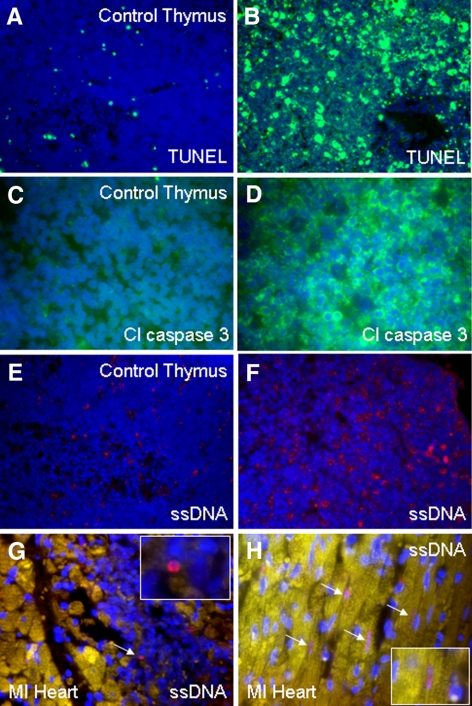
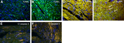
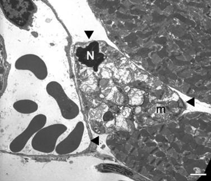
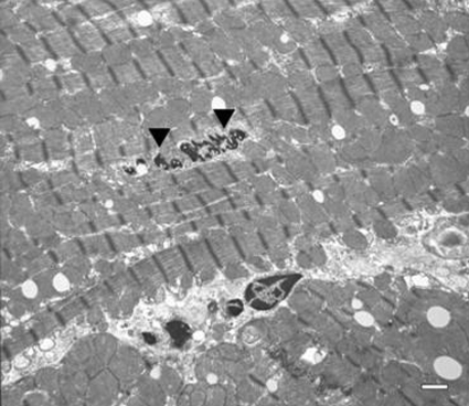
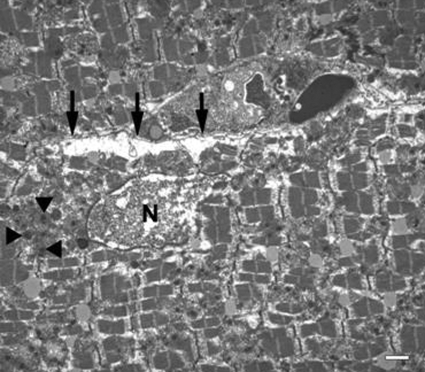
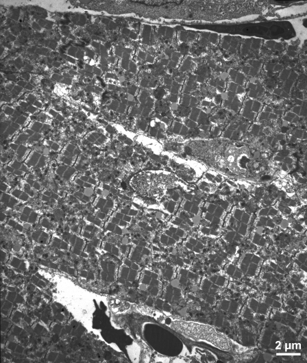
In view of the conventional wisdom in the cardiology literature that apoptosis is extensive early after myocardial ischemia, predicated largely from results with the TUNEL assay known to be nonspecific, this study was performed to delineate its extent with multiple assays and at multiple intervals. Coronary occlusion with and without subsequent revascularization was induced in 10-wk-old C57BL6 mice subjected to 1 or 4 h of transient ligation followed by 24 h of reperfusion, or 24 h persistent ligation. Apoptosis was quantified throughout the left ventricle immunohistochemically by assay of TUNEL, single-stranded DNA (ssDNA), and cleaved caspase 3; electron microscopy (EM); and activity assays of caspase 3 and 8. TUNEL staining was marked, but ssDNA and cleaved caspase 3 staining were significantly less (P<0.001 compared with TUNEL), and apoptosis defined by EM was virtually absent in all groups. Caspase 3 and caspase 8 activities per milligram protein were not significantly different from those in normal hearts. Only rare, potentially apoptotic cells were seen by EM in hearts from any group. Thus, the results with TUNEL were not specific, and the extent of apoptosis was markedly less than that predicated on the results with the TUNEL procedure. Apoptosis is de minimus early after transitory or persistent ischemia, though it is overestimated by TUNEL assays. Thus, antiapoptotic interventions per se are not likely to preserve substantial amounts of myocardium early after ischemic insults.


Figures

Figure 1.
Figure 1.
Confirmation of reperfusion with injection of Evans blue dye in the absence of ligation (A, B), after 4 h of persistent ligation (C, D), and after 4 h of transient ligation followed by reperfusion for 24 h (E, F). In each case, the heart was visualized before (left panels) and after injection of the dye. As can be seen, dye did not appear in nonreperfused regions (D) but did after 24 h of reperfusion (F). White regions in E and F are reflections from fat.
Figure 2.
Figure 2.
Left panels: thymus staining in nondexamethasone-treated mice for TUNEL (green) (A), cleaved caspase 3 (green) (C), and ssDNA (red) (E). Right panels: thymus staining in dexamethasone-treated mice showing significantly more positive staining for TUNEL (B), cleaved caspase 3 (D), and ssDNA (F). Rare positive ssDNA (red) staining is shown in heart from a mouse with MI in a lymphocyte (G) and in four cardiomyocytes (arrows) (H). Nuclei were stained with DAPI (blue). Yellow color is attributable to autofluorescence of tissue. Original views: ×40 (A, B, E, F); ×63 (C, D, G, H).
Figure 3.
Figure 3.
Sections from a mouse heart undergoing 1 h of transient ligation and 24 h of reperfusion (A, C, E) and from a mouse heart subjected to 24 h of persistent ligation (B, D, F) exhibit positive TUNEL staining (A, B) (green) that is much in excess and is not congruent with positive ssDNA (C, D) (pink) or positive cleaved caspase 3 staining (E, F) (green, none seen). Arrows indicate rare cells positive for ssDNA. Nuclei were stained with DAPI (blue). Yellow color is attributable to autofluorescence of tissue; mustard color is attributable to red blood cells. All panels ×40.
Figure 4.
Figure 4.
Transmission electron micrograph from the peri-infarct zone in the heart from an animal subjected to 4 h of transient ligation and 24 h of reperfusion. Cardiomyocyte in the middle (arrowheads) displays morphology potentially consistent with apoptosis in this cell type, including a shrunken nucleus (N) with condensed chromatin and abnormal swollen mitochondria (m) with “wrinkled” bodies. Scale bar = 2 μm.
Figure 5.
Figure 5.
Transmission electron micrograph from the ventricle of a control animal not subjected to surgery. Nucleus (arrowheads) in the center of the field displays marginated chromatin and atypical nuclear morphology. This is likely due to the plane of sectioning rather than apoptosis, since the rest of the cell evinces normal ultrastructural characteristics. Scale bar = 1 μm.
Figure 6.
Figure 6.
Transmission electron micrograph from the infarct zone in the heart from an animal subjected to 4 h transient coronary ligation and 24 h of reperfusion. Note the classic morphological features of necrosis rather than apoptosis: disrupted nucleus (N) with loss of nuclear content, electron-dense floccular deposits in mitochondria (arrowheads), and ruptured plasma membrane (arrows). Scale bar = 1 μm.
Figure 7.
Figure 7.
Another example of a transmission electron micrograph from an infarct zone from a mouse with 4-h transient coronary ligation and 24 h of reperfusion exhibiting a typical necrotic reaction. Scale bar = 2 μm.

References

    1. Sobel B E, Dauerman H L. The rationale underlying pharmacoinvasive therapy. Dauerman H L, Sobel BE, editors. New York: Taylor & Francis; Pharmacoinvasive Therapy in Acute Myocardial Infarction. 2005:3–31.
    1. Yellon D M, Hausenloy D J. Myocardial reperfusion injury. N Engl J Med. 2007;357:1121–1135. - PubMed
    1. Keeley E C, Boura J A, Grines C L. Primary angioplasty versus intravenous thrombolytic therapy for acute myocardial infarction: a quantitative review of 23 randomised trials. Lancet. 2003;361:13–20. - PubMed
    1. Xiang G, Schuster M D, Seki T, Witkowski P, Eshghi S, Itescu S. Downregulated expression of plasminogen activator inhibitor-1 augments myocardial neovascularization and reduces cardiomyocyte apoptosis after acute myocardial infarction. J Am Coll Cardiol. 2005;46:536–541. - PubMed
    1. Freude B, Masters T N, Robicsek F, Fokin A, Kostin S, Zimmermann R, Ullmann C, Lorenz-Meyer S, Schaper J. Apoptosis is initiated by myocardial ischemia and executed during reperfusion. J Mol Cell Cardiol. 2000;32:197–208. - PubMed

Publication types