. 2008 Dec 30;105(52):20641-6.
doi: 10.1073/pnas.0809680106. Epub 2008 Dec 18.

In vivo commitment and functional tissue regeneration using human embryonic stem cell-derived mesenchymal cells

Affiliations

In vivo commitment and functional tissue regeneration using human embryonic stem cell-derived mesenchymal cells

Nathaniel S Hwang et al. Proc Natl Acad Sci U S A. .

Abstract

Development of clinically relevant regenerative medicine therapies using human embryonic stem cells (hESCs) requires production of a simple and readily expandable cell population that can be directed to form functional 3D tissue in an in vivo environment. We describe an efficient derivation method and characterization of mesenchymal stem cells (MSCs) from hESCs (hESCd-MSCs) that have multilineage differentiation potential and are capable of producing fat, cartilage, and bone in vitro. Furthermore, we highlight their in vivo survival and commitment to the chondrogenic lineage in a microenvironment comprising chondrocyte-secreted morphogenetic factors and hydrogels. Normal cartilage architecture was established in rat osteochondral defects after treatment with chondrogenically-committed hESCd-MSCs. In view of the limited available cell sources for tissue engineering applications, these embryonic-derived cells show significant potential in musculoskeletal tissue regeneration applications.


Conflict of interest statement

The authors declare no conflict of interest.

Figures

Fig. 1.
Fig. 1.
Isolation and characterization of hESCd-MSCs. (A) EBs were formed from dissociated hESCs via liquid suspension methods for 10 days. EBs were then plated onto gelatin-coated (0.1% wt/vol) tissue culture plates, and migrating cells from EBs were subcultured with MSCM. (B) Inverted light microscopy of hESCd-MSCs pictures fibroblastic cell morphology. (Scale bar = 200 μm.) (C) Flow cytometry analysis of hESCd-MSCs indicates that these cells express MSC surface markers (at passage 7).
Fig. 2.
Fig. 2.
Developmental potential of hESCd-MSCs. (A) Chondrogenic differentiation in RGD-modified PEG hydrogels with TGF-β1 and ascorbic acid. Safranin-O staining (red) for negatively charged proteoglycans and type II collagen (type II collagen = red, nucleus = blue) in the pericellular signifies cartilaginous matrix production. RT-PCR confirmed chondrogenic differentiation of hESCd-MSCs with up-regulation of aggrecan and type II collagen. (B) The clusters of lipid-containing adipocytes were also detected by Oil red-O staining within 3 weeks of culture in adipogenic medium. Adipogenic differentiation was confirmed by the expression of αP2, lpl, and PPAR-γ by RT-PCR. (C) Bone-specific ALP was detected after 7 days, and mineralized deposits were evident with the alizarin red staining after 2 weeks of culture in osteo-inductive conditions. Osteogenic differentiation was confirmed by up-regulation of ALP, Cbfa1, and type I collagen by real-time PCR, and the expression of osteocalcin was confirmed by RT-PCR. (D) Gross image of in vivo-engineered bone tissues after 8 weeks of implantation. Microvascular perfusion and implant survival was assessed by luciferase detection of recombinant lentiviral vector-transduced hESCd-MSCs after 8 weeks of implantation. (E) H&E staining of the implanted scaffolds after 4 and 8 weeks. Histomophometric assessments showed increased bone area from 4 to 8 weeks (n = 4). UD, undifferentiated hESCd-MSCs; Dif, differentiated hESCd-MSCs.
Fig. 3.
Fig. 3.
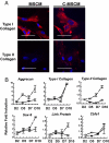
Effects of C-MSCM on hESCd-MSCs in vitro. (A) Type I collagen was detected in hESCd-MSCs, and upon expansion of C-MSCM, hESCd-MSCs displayed type II collagen. (Scale bars = 20 μm.) (B) Real-time PCR analysis of hESCd-MSCs in MSCM (○) or C-MSCM (□). Expression of cartilage-specific markers was analyzed for 10 days and normalized to day 1 control.
Fig. 4.
Fig. 4.
Chondrogenic differentiation potential of hESCd-MSCs in vivo. Control medium-expanded or conditioned medium-expanded hESCd-MSCs were encapsulated in PEGDA hydrogels and subsequently implanted into the s.c. space of an athymic mouse. (A) Gross image of hydrogels after 12 weeks of implantation. Safranin-O staining and immunostaining for extracellular matrix proteins produced by hESCd-MSCs after 12 weeks of implantation indicated that unstimulated hESCd-MSCs lacked differentiation potential. (B and C) Conditioned medium-expanded hESCd-MSCs produced cartilage-like tissue as indicated by Safranin-O staining (B) and immunostaining for cartilage-specific extracellular matrix molecules (C). (D–G) C-MSCM-expanded hESCd-MSCs resulted in engineered cartilage with enhanced mechanical strength and higher ECM contents. *, P < 0.05; **, P < 0.01. (Scale bars = 100 μm.)
Fig. 5.
Fig. 5.
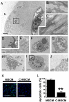
Electron micrographs demonstrated marked differences in cell size, shape, and organization of pericellular extracellular matrix after 12 weeks in vivo. (A–G) hESCd-MSCs expanded in C-MSCM maintained round chondrocyte morphology with the collagen matrix assembled in the pericellular region. (H–J) However, irregular cellular morphology reminiscent of apoptotic cells was observed in the hESCd-MSCs expanded in control medium (H) without any detectable extracellular matrix in the pericellular region (I–J) (n = nucleus, h = hydrogel). (K and L) TUNEL assay confirmed the presence of more apoptotic cells in the hESCd-MSCs expanded in control medium compared with that of C-MSCM. (Scale bars: A, F–I = 6 μm; B, D, E, J = 1 μm; C = 500 nm; K = 50 μm.)
Fig. 6.
Fig. 6.
Osteochondral defects created in a rat knee (femorapatellar groove) repaired with hESCd-MSC pellets 8 weeks after transplantation. (A) Defects implanted with cell pellets generated articular cartilage. (B) Histomorphometric analysis of quantified Safranin-O positive area (n = 6). *, P < 0.05; **, P < 0.01.

References

    1. Lerou PH, Daley GQ. Therapeutic potential of embryonic stem cells. Blood Rev. 2005;19:321–331. - PubMed
    1. Thomson JA, et al. Embryonic stem cell lines derived from human blastocysts. Science. 1998;282:1145–1147. - PubMed
    1. Takahashi K, Yamanaka S. Induction of pluripotent stem cells from mouse embryonic and adult fibroblast cultures by defined factors. Cell. 2006;126:663–676. - PubMed
    1. Park IH, et al. Disease-specific induced pluripotent stem cells. Cell. 2008;134:877–886. - PMC - PubMed
    1. Barberi T, Willis LM, Socci ND, Studer L. Derivation of multipotent mesenchymal precursors from human embryonic stem cells. PLoS Med. 2005;2:e161. - PMC - PubMed

Publication types

LinkOut - more resources