. 2008 Dec 30;105(52):20671-6.
doi: 10.1073/pnas.0810463106. Epub 2008 Dec 18.

The crystal structure of the second Z-DNA binding domain of human DAI (ZBP1) in complex with Z-DNA reveals an unusual binding mode to Z-DNA

Affiliations

The crystal structure of the second Z-DNA binding domain of human DAI (ZBP1) in complex with Z-DNA reveals an unusual binding mode to Z-DNA

Sung Chul Ha et al. Proc Natl Acad Sci U S A. .

Abstract

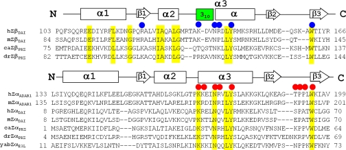
Mammalian DAI (DNA-dependent activator of IFN-regulatory factors), an activator of the innate immune response, senses cytosolic DNA by using 2 N-terminal Z-DNA binding domains (ZBDs) and a third putative DNA binding domain located next to the second ZBD. Compared with other previously known ZBDs, the second ZBD of human DAI (hZbeta(DAI)) shows significant variation in the sequence of the residues that are essential for DNA binding. In this article, the crystal structure of the hZbeta(DAI)/Z-DNA complex reveals that hZbeta(DAI) has a similar fold to that of other ZBDs, but adopts an unusual binding mode for recognition of Z-DNA. A residue in the first beta-strand rather than residues in the beta-loop contributes to DNA binding, and part of the (alpha3) recognition helix adopts a 3(10) helix conformation. The role of each residue that makes contact with DNA was confirmed by mutational analysis. The 2 ZBDs of DAI can together bind to DNA and both are necessary for full B-to-Z conversion. It is possible that binding 2 DAIs to 1 dsDNA brings about dimerization of DAI that might facilitate DNA-mediated innate immune activation.


Conflict of interest statement

The authors declare no conflict of interest.

Figures

Fig. 1.
Fig. 1.
Multiple sequence alignments of the first ZBDs (Zα) and the second ZBDs (Zβ). Zαs include human ADAR1 (hZαADAR1), mouse ADAR1 (mZαADAR1), human DAI (hZαDAI), mouse DAI (mZαDAI), goldfish PKZ (caZαPKZ), zebrafish PKZ (drZαPKZ), and Yaba-like disease poxvirus E3L (yabZαE3L). Zβs include human DAI (hZβDAI), mouse DAI (mZβDAI), goldfish PKZ (caZβPKZ), and zebrafish PKZ (drZβPKZ). The secondary structures of hZβDAI and hZαADAR1 are shown by a rectangle indicating a helix or an arrow for a β-strand. The 310 helix is shown in green. Residues of hZβDAI and hZαADAR1 involved in DNA interactions are marked by blue and red circles, respectively. Highly conserved residues are highlighted in yellow. The residue numbers of both N and C termini are shown.
Fig. 2.
Fig. 2.
Structural comparison of hZβDAI and hZαADAR1 and their interactions with Z-DNA. (A) Overall structure of the hZβDAI/Z-DNA complex. The protein and DNA are drawn as a ribbon diagram and stick model, respectively. The N and C termini, the secondary structure elements of hZβDAI, and 5′ and 3′ of DNA are labeled. (B) The structural overlap of hZβDAI and hZαADAR1 near the Z-DNA binding site. The ribbon diagrams of hZβDAI (blue) and hZαADAR1 (green) are overlapped, and the stick model of a double-strand Z-DNA is drawn in brown and gray. Structural deviation near α3 and the β-wing are labeled and marked by red circles. (C) Stereoviews of the protein–DNA interfaces of hZβDAI (chain A)/Z-DNA (Left) and hZβDAI(Chain B)/Z-DNA (Right). The Cα chains of the ZBDs are colored sky blue, and the residues involved in DNA contact are depicted in sky blue stick models. The Z-DNA backbone is drawn as a red stick model, and bases are drawn as gray stick models. Water molecules are shown in green. Hydrogen bonds are drawn as dashed lines. (D) Schematic diagrams of the protein–Z-DNA interactions in hZβDAI (Left) and hZαADAR1 (Right). Hydrogen bonds and van der Waals contacts are represented by dashed black lines and pink lines, respectively. The CH–π interaction between the conserved Tyr and the C8 of a syn-guanine is indicated by black circles, and waters are shown by green circles. The protein–DNA interactions in hZαADAR1 are identical on both sides of the Z-DNA, thus only 1 side is shown.
Fig. 3.
Fig. 3.
The Z-DNA binding mode of hZβDAI. (A) Z-DNA recognition by R124 of hZβDAI. A magnified view near the wing region is drawn for the interfaces between hZβDAI and Z-DNA [chains A and C (Left) and chains B and D (Right)]. A σ-weighted 2 FoFc omit map contoured at 1.2 σ was generated by omitting R124, R160, and the waters involved in the R124-mediated interaction. Possible hydrogen bonds among R124, water and the phosphate group of G2 are shown by dashed lines, and their distances are indicated in Å. (B) Surface charge distributions of the chain A (Left) and chain B (Right) viewed along the DNA binding cleft. The red and blue areas represent negatively and positively charged surfaces, respectively. Z-DNA chains are shown as stick models, and R124, K160 and each nucleotide are labeled.
Fig. 4.
Fig. 4.
B-to-Z-DNA conversion activity of hZβDAI and hZαβDAI. (A) The Z-converting activities were estimated by monitoring the CD signal at 255 nm for 40 min by using 60 μg/ml of double-stranded (dCdG)6. The molar ratio of hZβDAI or its alanine mutants to ds(dCdG)6 was 4:1. D139A was randomly chosen as a negative control. (B) WT hZαβDAI and 2 tyrosine mutants, hZαβDAI Y50A and hZαβDAI Y145A, were mixed with 15 μM double-stranded (dCdG)6, at 2:1 and 4:1 molar ratios, and their CD signals were monitored at 255 nm for 60 min. As a control, the Z-DNA converting activity of a single ZBD (hZαADAR1) at a 4:1 molar ratio to DNA was also measured under the same condition.

Similar articles

Cited by

References

    1. Eda K, Akira S. Toll-like receptors in innate immunity. Int Immunol. 2005;17:1–14. - PubMed
    1. Ishii KJ, Akira S. Innate immune recognition of, and regulation by, DNA. Trends Immunol. 2006;27:525–532. - PubMed
    1. Ishii KJ, et al. A Toll-like receptor-independent antiviral response induced by double-stranded B-form DNA. Nat Immunol. 2006;7:40–48. - PubMed
    1. Takaoka A, et al. DAI(DLM-1/ZBP1) is a cytosolic DNA sensor and an activator of innate immune response. Nature. 2007;448:501–504. - PubMed
    1. Wang Z, et al. Regulation of innate immune responses by DAI (DLM-1/ZBP1) and other DNA-sensing molecules. Proc Natl Acad Sci USA. 2008;105:5477–5482. - PMC - PubMed

Publication types

Associated data