. 2009 Jan 1;23(1):54-66.
doi: 10.1101/gad.1717309. Epub 2008 Dec 18.

SMD and NMD are competitive pathways that contribute to myogenesis: effects on PAX3 and myogenin mRNAs

Affiliations

SMD and NMD are competitive pathways that contribute to myogenesis: effects on PAX3 and myogenin mRNAs

Chenguang Gong et al. Genes Dev. .

Abstract

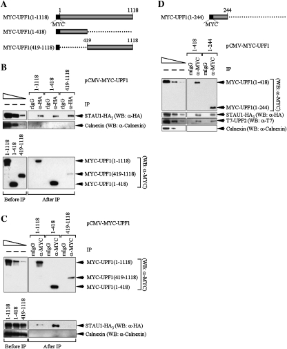
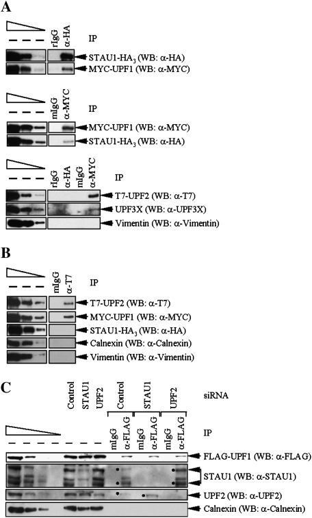
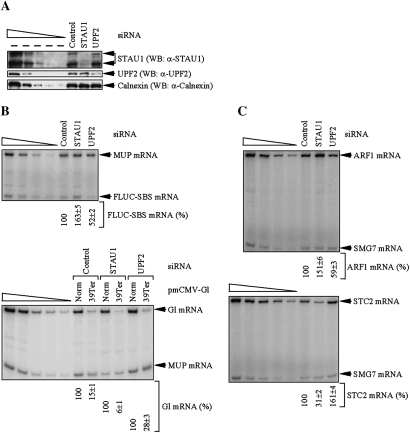
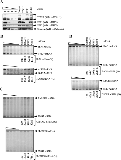
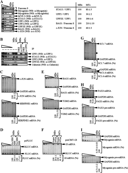
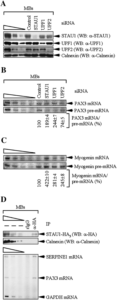
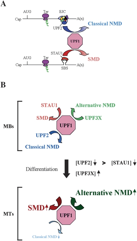
UPF1 functions in both Staufen 1 (STAU1)-mediated mRNA decay (SMD) and nonsense-mediated mRNA decay (NMD), which we show here are competitive pathways. STAU1- and UPF2-binding sites within UPF1 overlap so that STAU1 and UPF2 binding to UPF1 appear to be mutually exclusive. Furthermore, down-regulating the cellular abundance of STAU1, which inhibits SMD, increases the efficiency of NMD, whereas down-regulating the cellular abundance of UPF2, which inhibits NMD, increases the efficiency of SMD. Competition under physiological conditions is exemplified during the differentiation of C2C12 myoblasts to myotubes: The efficiency of SMD increases and the efficiency of NMD decreases, consistent with our finding that more STAU1 but less UPF2 bind UPF1 in myotubes compared with myoblasts. Moreover, an increase in the cellular level of UPF3X during myogenesis results in an increase in the efficiency of an alternative NMD pathway that, unlike classical NMD, is largely insensitive to UPF2 down-regulation. We discuss the remarkable balance between SMD and the two types of NMD in view of data indicating that PAX3 mRNA is an SMD target whose decay promotes myogenesis whereas myogenin mRNA is a classical NMD target encoding a protein required for myogenesis.


Figures

Figure 1.
Figure 1.
Amino acids 1–244 of UPF1 interact with STAU1 or UPF2. (A) Diagram of full-length MYC-UPF1 (1–1118) and two deletion variants. (B) Lysates of Cos cells (3 × 10⁷ cells per 150-mm dish) that had been transiently transfected with 10 μg of pSTAU1-HA3 and 5 μg of pCMV-MYC-UPF1(1–1118), 4 μg of pCMV-MYC-UPF1(1–418), or 15 μg of pCMV-MYC-UPF1(419–1118) (plus a variable amount of pUC19 for a total of 25 μg of transfecting DNA) were immunoprecipitated using anti(α)-HA or, to control for nonspecific IP, rIgG. Equivalent amounts of immunoprecipitated proteins were then analyzed by Western blotting (WB) before (−) IP (refer to the left-most lane when dilutions were analyzed) or after IP using anti-HA, anti-MYC or, as a control for IP specificity, anti-Calnexin. The top three left-most lanes represent threefold dilutions of protein before (−) IP and demonstrate that the conditions used for Western blotting are semiquantitative. (C) As in B, except lysates were immunoprecipitated using anti-MYC or, to control for nonspecific IP, mIgG. (D, top) Diagram of MYC-UPF1(1–244). (D, bottom) As in C, except cells were transfected with 10 μg of pSTAU1-HA3, 10 μg of pCI-NEO-T7-UPF2, and 5 μg of either pCMV-MYC-UPF1(1–418) or pCMV-MYC-UPF1(1–244). Results are representative of five independently performed experiments.
Figure 2.
Figure 2.
Evidence that STAU1 and UPF2 compete for binding to UPF1. (A) Cos cells (3 × 10⁷) were transiently transfected with 10 μg of pSTAU1-HA3, 10 μg of pCMV-MYC-UPF1, and 10 μg of pCI-NEO-T7-UPF2. Protein was purified before (−) and after IP using anti(α)-HA or, to control for nonspecific IP, rIgG (top); anti-MYC or, to control for nonspecific IP, mgG (middle); or all four antibodies (bottom). Equivalent amounts of immunoprecipitated proteins were analyzed by Western blotting (WB) using antibody to the appropriate epitope tag or Vimentin. (B) Cos cells were transiently transfected as in A, and protein was purified before and after IP using anti-T7 or, to control for nonspecific IP, mIgG. Western blotting was performed using the antibody to the appropriate epitope, Calnexin or Vimentin. (C) HeLa cells (3 × 10⁷) that stably express Flag-UPF1 were transiently transfected with 100 nM of STAU1 siRNA, UPF2 siRNA, or a nonspecific Control siRNA. Three days later, protein was purified before and after IP using anti-Flag or, as a control, mIgG that had been covalently conjugated to an agarose affinity gel. Protein was then analyzed by Western blotting using anti-Flag, anti-STAU1, anti-UPF2, or anti-Calnexin. Dots specify the two STAU1 isoforms or UPF2. For each panel, protein from the same number of cells was analyzed in the left-most lane of the dilution standards and in each IP.
Figure 3.
Figure 3.
Decreasing the cellular abundance of STAU1 increases the efficiency of NMD, and decreasing the cellular abundance of UPF2 increases the efficiency of SMD. HeLa cells (2 × 10⁶) were transiently transfected with the 100 nM of STAU1 siRNA, UPF2 siRNA, or Control siRNA. Two days later, cells were retransfected with 0.3 μg of the specified pmCMV-Gl test plasmid, 0.3 μg of the specified pcFLUC test plasmid, and 0.2 μg of the phCMV-MUP reference plasmid. After an additional 12 h, protein and RNA were analyzed. (A) Western blotting (WB) using anti(α)-STAU1, anti-UPF2, or anti-Calnexin. (B) RT–PCR analysis, where the level of FLUC-SBS or Gl mRNA was normalized to the level of MUP mRNA, and the normalized level of FLUC-SBS mRNA in the presence of Control siRNA or Gl Norm mRNA in the presence of each siRNA was defined as 100. (C) RT–PCR analysis, where the level of ARF1 or STC2 mRNA was normalized to the level of SMG7 mRNA, and the normalized value in the presence of Control siRNA was defined as 100. All results are representative of three independently performed experiments that did not vary by more than the amount shown.
Figure 4.
Figure 4.
HeLa cell transcripts down-regulated upon UPF2 down-regulation include SMD targets. HeLa cells (2 × 10⁷) were transiently transfected with 100 nM of the specified siRNA and harvested after an additional 48 h. (A) Western blotting (WB) demonstrated the degree of siRNA-mediated down-regulation of STAU1, UPF1, or UPF2. (B) RT–PCR analysis of IL7R or c-JUN mRNA after normalization to the level of SMG7 mRNA, where the level in the presence of Control siRNA was defined as 100. (C) As in B, except that AMIGO2 or FLJ21870 mRNA was analyzed. (D) As in B, except that BAG1 or DSCR1 mRNA was analyzed. All results are representative of three independently performed experiments that did not vary by more than the amount shown.
Figure 5.
Figure 5.
Myogenic differentiation of C2C12 cells is accompanied by an increase in the efficiency of SMD, a decrease in the efficiency of classical NMD, and an increase in the efficiency of alternative NMD. MBs or MTs (2 × 10⁷) were transiently transfected with three plasmids: 3 μg of pmCMV-Gl Norm or pmCMV-Gl Ter, 12 μg of pFLUC-No SBS or pFLUC-SBS, and 3 μg of pRLUC. Two days later, cells were harvested. (A, left) Western blotting (WB) of myogenin, myoglobin, MYF-5, STAU1, UPF1, UPF2, UPF3X, BAG1, or c-JUN. Ponceau S staining was used as a loading control. (A, right) Quantitation of the level of STAU1, UPF2, or UPF3X relative to UPF1, or BAG1 or c-JUN relative to Ponceau S staining, in MBs and MTs. Each level in MBs was defined as 100. (B) Western blotting of specified proteins before (−) or after IP using anti-UPF1 or, to control for nonspecific IP, normal rabbit serum (NRS). (C, top and bottom) RT–PCR analysis of c-JUN or SERPINE1 mRNA after normalization to the level of GAPDH mRNA, where each normalized level in MBs was defined as 100. (D) The level of FLUC-No SBS or FLUC-SBS mRNA was normalized to the level of RLUC mRNA, and the normalized level of FLUC-No SBS in MBs or MTs was defined as 100. (E, top and bottom) RT–PCR analysis of BAG1 or TGM2 mRNA after normalization to the level of GAPDH mRNA, where each normalized level in MBs was defined as 100. (F) The level of PTC-free (Norm) or PTC-containing (Ter) Gl mRNA was normalized to the level of RLUC mRNA, and the normalized level of G1 Norm mRNA in MBs or MTs was defined as 100. (G, top and bottom) RT–PCR analysis of SC1.7 or SC1.6 mRNA after normalization to the level of GAPDH mRNA, where each normalized level in MBs was defined as 100 (asterisk denotes a nonspecific RT–PCR product that does not interfere with the analysis). (H, top and bottom) As in G. However, PAX3 mRNA or pre-mRNA was analyzed. (I, top and bottom) As in G. However, myogenin mRNA or pre-mRNA was analyzed. All results are representative of at least three independently performed experiments that did not vary by more than the amount shown.
Figure 6.
Figure 6.
Evidence that PAX3 mRNA is an SMD target and myogenin mRNA is an NMD target. (A–C) MBs (1 × 10⁶) were transiently transfected with 100 nM of the specified siRNA and harvested after an additional 48 h. (A) Western blotting (WB) demonstrated the degree of siRNA-mediated down-regulation of STAU1, UPF1, or UPF2. Notably, mouse STAU1, unlike human STAU1, is detected as a 55-kDa isoform but not a 63-kDa isoform. (B) RT–PCR analysis of PAX3 mRNA after normalization to the level of PAX3 pre-mRNA, where the level in the presence of Control siRNA was defined as 100. Notably, equal amounts of total cell RNA rather than equal amounts of PAX3 pre-mRNA were analyzed in each lane. (C) As in B, except that myogenin mRNA or pre-mRNA was analyzed. (D) MBs (2 × 10⁷) were transiently transfected with 10 μg of pSTAU1-HA3. Two days later, cells were harvested, treated with formaldehyde to cross-link protein and RNA, and lysed using sonication. Lysates were immunoprecipitated using anti-HA or, to control for nonspecific IP, rIgG, and cross-links were reversed using heat. Protein was analyzed by Western blotting using anti-HA or anti-Calnexin. RNA was analyzed by RT–PCR for SERPINE1, PAX3, or GAPDH mRNA. All results are representative of at least three independently performed experiments that did not vary by more than the amount shown.
Figure 7.
Figure 7.
Model for competition between SMD and NMD. (A) UPF2, which is an EJC constituent and functions in the classical NMD pathway, and STAU1, which is an RNA-binding protein that functions in the SMD pathway, compete for binding to UPF1 and, thus, the recruitment of UPF1 to mRNA. UPF1 functions in both pathways to elicit mRNA decay when translation terminates sufficiently upstream of an EJC, in the case of NMD, or a SBS, in the case of SMD. Ter can be either a premature or normal termination codon. (B) As a consequence of C2C12 cell differentiation from MBs to MTs, the efficiency of SMD increases, the efficiency of classical (i.e., UPF2 siRNA-sensitive) NMD decreases, and the efficiency of an alternative NMD pathway that depends on UPF3X but not appreciably UPF2 increases. During myogenesis, a larger decrease in the abundance of UPF2, which drops to almost undetectable levels, relative to STAU1 permits STAU1 to out-compete UPF2 for binding to UPF1, and an approximately fourfold increase in the level of UPF3X supports an increase in the efficiency of the alternative NMD pathway.

References

    1. Behm-Ansmant I., Kashima I., Rehwinkel J., Sauliere J., Wittkopp N., Izaurralde E. mRNA quality control: An ancient machinery recognizes and degrades mRNAs with nonsense codons. FEBS Lett. 2007;581:2845–2853. - PubMed
    1. Belgrader P., Maquat L.E. Nonsense but not missense mutations can decrease the abundance of nuclear mRNA for the mouse major urinary protein, while both types of mutations can facilitate exon skipping. Mol. Cell. Biol. 1994;14:6326–6336. - PMC - PubMed
    1. Berger F.G., Szoka P. Biosynthesis of the major urinary proteins in mouse liver: A biochemical genetic study. Biochem. Genet. 1981;19:1261–1273. - PubMed
    1. Bhattacharya A., Czaplinski K., Trifillis P., He F., Jacobson A., Peltz S.W. Characterization of the biochemical properties of the human Upf1 gene product that is involved in nonsense-mediated mRNA decay. RNA. 2000;6:1226–1235. - PMC - PubMed
    1. Blais A., Tsikitis M., Acosta-Alvear D., Sharan R., Kluger Y., Dynlacht B.D. An initial blueprint for myogenic differentiation. Genes & Dev. 2005;19:553–569. - PMC - PubMed

Publication types

MeSH terms