. 2009 Jan;174(1):330-42.
doi: 10.2353/ajpath.2009.080385. Epub 2008 Dec 18.

Valproate attenuates accelerated atherosclerosis in hyperglycemic apoE-deficient mice: evidence in support of a role for endoplasmic reticulum stress and glycogen synthase kinase-3 in lesion development and hepatic steatosis

Affiliations

Valproate attenuates accelerated atherosclerosis in hyperglycemic apoE-deficient mice: evidence in support of a role for endoplasmic reticulum stress and glycogen synthase kinase-3 in lesion development and hepatic steatosis

Anna J Bowes et al. Am J Pathol. 2009 Jan.

Abstract

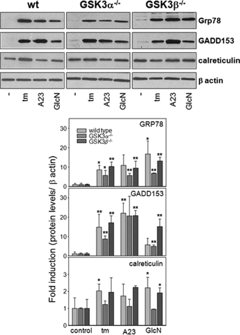
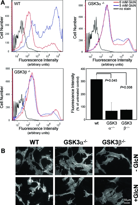
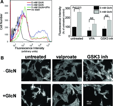
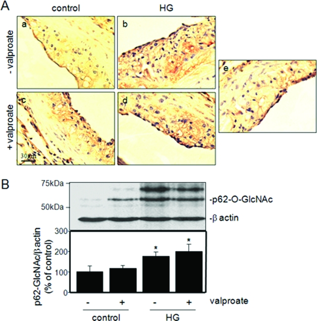
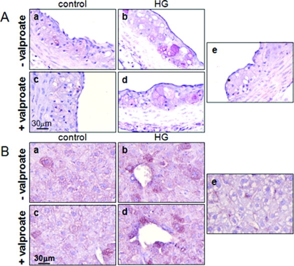
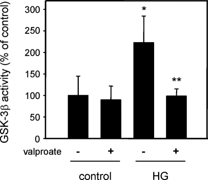
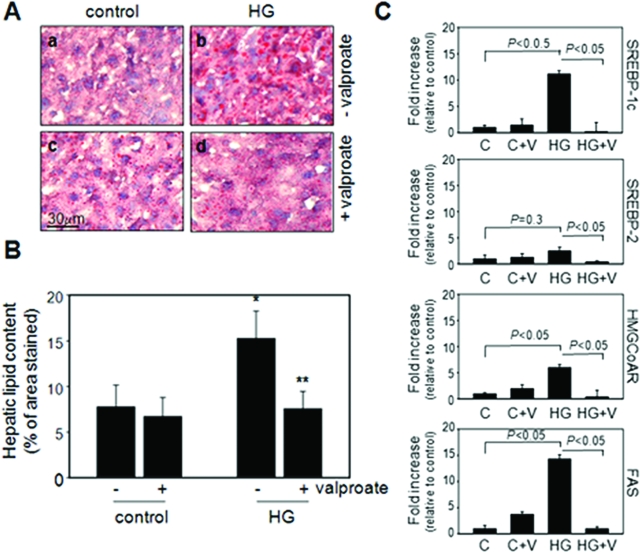
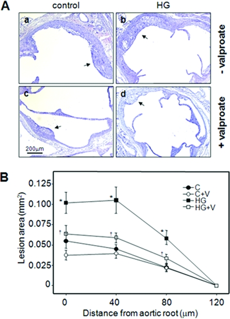
We have previously shown that glucosamine promotes endoplasmic reticulum (ER) stress in vascular cells leading to both inflammation and lipid accumulation--the hallmark features of atherosclerosis. Pretreatment with glycogen synthase kinase (GSK)-3 inhibitors protects cultured cells from ER stress-induced dysfunction. Here we evaluate the potential role of GSK-3 on the pro-atherogenic effects of hyperglycemia and ER stress. We show that GSK-3-deficient mouse embryonic fibroblasts do not accumulate unesterified cholesterol under conditions of ER stress. Furthermore, GSK-3 inhibitors, including valproate, attenuate ER stress-induced unesterified cholesterol accumulation in wild-type mouse embryonic fibroblasts. In vivo we show that hyperglycemic apoE-deficient mice have accelerated atherogenesis at the aortic root compared with normoglycemic control mice. Mice fed a diet supplemented with 625 mg/kg valproate have significantly reduced lesion volume relative to nonsupplemented controls. Valproate supplementation has no apparent effect on the plasma levels of either glucose or lipids or on the expression of diagnostic markers of ER stress in the lesion. Significant reductions were observed in total hepatic lipids (>50.4%) and hepatic GSK-3beta activity (>55.8%) in mice fed the valproate diet. In conclusion, dietary supplementation with low levels of valproate significantly attenuates atherogenesis in hyperglycemic apoE-deficient mice. The in vivo anti-atherogenic effects of valproate are consistent with its ability to inhibit GSK-3 and interfere with pro-atherogenic ER stress signaling pathways in vitro.


Figures

Figure 1
Figure 1
The UPR in GSK-3-deficient MEFs. A: Wild-type, GSK-3α−/−, and GSK-3β−/− MEFs were challenged with ER stress-inducing agents including tunicamycin, A23187, or glucosamine for 18 hours as indicated. Total protein lysates were resolved by sodium dodecyl sulfate-polyacrylamide gel electrophoresis, transferred to nitrocellulose membranes, and immunostained with antibodies against GRP78/BiP, GADD153, calreticulin, or β-actin. B: The amount of each protein normalized to β-actin and averaged over three independent experiments was quantified by densitometry. *P < 0.05, **P < 0.01 relative to untreated controls.
Figure 2
Figure 2
The effect of GSK-3 deficiency on glucosamine-induced lipid accumulation. Wild-type, GSK-3α−/−, and GSK-3β−/− MEFs were challenged with 0 or 5 mmol/L glucosamine for 18 hours. Cells were stained with filipin and fluorescence was examined by FACS (A) and fluorescence microscopy (B). For FACS, >10,000 cells were analyzed from each treatment group using excitation wavelengths of 350 to 370 nm and emission detection at 420 to 460 nm.
Figure 3
Figure 3
The effect of GSK-3 inhibition on glucosamine-induced lipid accumulation. Wild-type MEFs were challenged with 0 or 5 mmol/L glucosamine in the presence of valproate or 200 μmol/L GSK-3 inhibitor II. Cells were stained with filipin and analyzed by FACS (A) and fluorescence microscopy (B) as described in Figure 2.
Figure 4
Figure 4
Effect of valproate supplementation on intracellular O-linked glycosylation. A: Representative sections of aortic root of control female normoglycemic and hyperglycemic apoE−/− mice fed control diet (a, b) or control diet supplemented with sodium valproate (c, d) were stained with an antibody against O-linked N-acetylglucosamine (CTD110.6) or pre-immune IgG (e). B: Total protein lysates from liver were analyzed by immunoblot analysis using the RL-2 antibody. The relative amounts of the O-glycosylated nuclear pore protein, p62 (p62-O-GlcNAc), an established marker of intracellular glucosamine levels, were quantified and plotted for each group. n = 3 to 6 per group; *P < 0.05 relative to controls.
Figure 5
Figure 5
Effect of valproate on markers of ER stress. A: Representative sections of aortic root from each of the indicated treatment groups were immunostained with antibodies against phosphorylated-PERK (a–d) or pre-immune IgG (e). B: Sections of liver from each of the treatment groups were immunostained with anti-KDEL antibody (a–d) or pre-immune IgG (e) as indicated.
Figure 6
Figure 6
Effect of valproate supplementation on hepatic GSK-3 activity. GSK-3β was immunoprecipitated from protein lysates prepared from the livers of normoglycemic (control) and hyperglycemic (HG) mice fed control or valproate-supplemented diet. GSK-3β kinase activity was determined as previously described. n = 3, *P < 0.05 relative to nonsupplemented control. **P < 0.05 relative to nonsupplemented HG.
Figure 7
Figure 7
Effect of valproate supplementation on hepatic steatosis. A: Representative sections of liver from control or hyperglycemic apoE−/− mice fed control diet or valproate-supplemented diet, as indicated, were stained with Oil Red O. B: Quantification of Oil Red O staining in representative cross sections. n = 3 per group. C: Quantification of mRNA levels using quantitative RT-PCR in the liver lysates from indicated groups. *P < 0.05 relative to control mice, **P < 0.05 relative to mice from the same treatment group without valproate supplementation.
Figure 8
Figure 8
Effect of valproate supplementation on atherosclerotic plaque area in hyperglycemic apoE-deficient mice. A: Representative H&E-stained aortic root cross sections from 15-week-old control female normoglycemic and hyperglycemic apoE−/− mice fed control diet (a, b) or control diet supplemented with sodium valproate (c, d) as indicated. B: Lesion areas were determined at the aortic root (0 μm) and at 40-μm intervals in aortae isolated from control (C) or hyperglycemic (HG) mice fed control diet or control diet supplemented with sodium valproate (+V). n = 9 to 10 per group; *P < 0.05, relative to control mice; P < 0.05, relative to similar treatment group in the absence of valproate supplementation. Arrows indicate atherosclerotic lesions.

References

    1. Haffner SM, Lehto S, Ronnemaa T, Pyorala K, Laakso M. Mortality from coronary heart disease in subjects with type2 diabetes and in nondiabetic subjects with and without prior myocardial infarction. N Engl J Med. 1998;339:229–234. - PubMed
    1. Nathan DM, Lachin J, Cleary P, Orchard T, Brillon DJ, Backlund JY, O'Leary DH, Genuth S, the Diabetes Control and Complications Trial Epidemiology of Diabetes. N Engl J Med. 2003;348:2294–2303. - PMC - PubMed
    1. Turner RC, Holman RR, Cull CA, Stratton IM, Matthews DR, Frighi V, Manley SE, Neil A, McElroy H, Wright D, Kohner E, Fox C, Hadden D, the UK Prospective Diabetes Lancet. 1998;352:837–853.
    1. Standl E, Balletshofer B, Dahl B, Weichenhain B, Steigler H, Hormann A, Holl R. Predictors of 10-year macrovascular and overall mortality in patients with NIDDM: the Munich General Practitioner Project. Diabetologia. 1996;39:1540–1545. - PubMed
    1. Marciniak SJ, Ron D. Endoplasmic reticulum stress signaling in disease. Physiol Rev. 2006;86:1133–1149. - PubMed

Publication types

MeSH terms