. 2009 Jan;174(1):44-53.
doi: 10.2353/ajpath.2009.080505. Epub 2008 Dec 18.

Adipose tissue sensitivity to radiation exposure

Affiliations

Adipose tissue sensitivity to radiation exposure

Sandrine Poglio et al. Am J Pathol. 2009 Jan.

Abstract

Treatment of cancer using radiation can be significantly compromised by the development of severe acute and late damage to normal tissue. Treatments that either reduce the risk and severity of damage or that facilitate the healing of radiation injuries are being developed, including autologous adipose tissue grafts to repair tissue defects or involutional disorders that result from tumor resection. Adipose tissue is specialized in energy storage and contains different cell types, including preadipocytes, which could be used for autologous transplantation. It has long been considered a poorly proliferative connective tissue; however, the acute effects of ionizing radiation on adipose tissue have not been investigated. Therefore, the aim of this study was to characterize the alterations induced in adipose tissue by total body irradiation. A severe decrease in proliferating cells, as well as a significant increase in apoptotic cells, was observed in vivo in inguinal fat pads following irradiation. Additionally, irradiation altered the hematopoietic population. Decreases in the proliferation and differentiation capacities of non-hematopoietic progenitors were also observed following irradiation. Together, these data demonstrate that subcutaneous adipose tissue is very sensitive to irradiation, leading to a profound alteration of its developmental potential. This damage could also alter the reconstructive properties of adipose tissue and, therefore, calls into question its use in autologous fat transfer following radiotherapy.


Figures

Figure 1
Figure 1
Morphological characterization of subcutaneous adipose tissue after irradiation. May-Grunwald Giemsa staining was performed on white adipose tissue isolated from control mice (A) or mice 7 days after sublethal (B) or lethal total body irradiation (C). Mature adipocytes diameter (D) was measured on adipose tissue slides. Three populations have been distinguished according to their diameter: less than 50 μm; from 50 to 100 μm; and greater than 100 μm. The quantification was performed in control, sublethally (7Gy), and lethally (10Gy) irradiated mice. *P < 0.05.
Figure 2
Figure 2
Effect of irradiation on proliferation and apoptosis in inguinal adipose tissue. Ki-67 staining (A–C) or TUNEL assay (D–F) were performed on paraffin embedded adipose tissue isolated from control mice (A, D) or mice 7 days after sublethal (B, E) or lethal (C, F) irradiation. Ki-67 staining was revealed by AEC, and TUNEL positive cells were revealed by DAB (dark nuclei, black arrows). In both cases, slides were counterstained with hematoxylin to visualize negative nuclei (blue arrows). Red arrows point on mature adipocytes positive for TUNEL.
Figure 3
Figure 3
Effect of irradiation on radical oxidative stress in inguinal adipose tissue. NADPH oxidase and MnSOD gene expression were quantified by quantitative reverse transcription PCR (A), and aconitase activity (B) was measured in subcutaneous fat pad removed from control (white bars), sublethally (gray bars) or lethally (black bars) irradiated mice. *P < 0.05; **P < 0.01; n = 3.
Figure 4
Figure 4
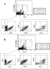
Effect of irradiation on SVF cell subsets. Flow cytometry was performed to compare the percentage of cells expressing the pan-hematopoietic cell surface marker CD45 in combination with specific markers for endothelial cells (CD31/CD34) or preadipocytes (CD34/CD90/ScaI) in total SVF from control (A) or lethally irradiated (B) mice. Dot plots were representative of 8 to 10 experiments.
Figure 5
Figure 5
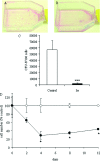
Effect of irradiation on cell proliferation. Cells isolated from inguinal adipose tissue from control (white symbols) or lethally irradiated (black symbols) mice were cultured either in clonal conditions to obtain CFU-f (A–C) or in liquid medium (D) as described in Material and Methods. For CFU-f quantification, Giemsa staining was performed in the flask of control cells (A) or cells isolated from irradiated mice (B), and the number of CFU-f was quantified (C). For culture in liquid medium (D), at indicated time, cells were trypsinized and counted. The y axis shows cell number expressed as a percentage of control values. The results represented the mean ± SEM of 4 to 6 cultures. *P < 0.05; ***P < 0.001.
Figure 6
Figure 6
Adipocyte differentiation of SVF cells isolated from inguinal adipose tissue of control and lethally irradiated mice. Phase contrast pictures of differentiated cultures obtained from control (A) and lethally irradiated (B) mice. Quantification of triglyceride content (C) in each culture was performed 8 days after induction of adipocyte differentiation, and was expressed as fold increased in differentiated versus confluent cultures. D: Adipogenic clone number in methylcellulose cultures were counted and expressed per well. White bar = control mice; black bars = irradiated mice. The results are expressed as the mean ± sem of 4–6 cultures. *P < 0.05.

Similar articles

Cited by

References

    1. Barcellos-Hoff MH, Park C, Wright EG. Radiation and the microenvironment- tumorigenesis and therapy. Nat Rev Cancer. 2005;5:867–875. - PubMed
    1. Bentzen SM. Preventing or reducing late side effects of radiation therapy: radiobiology meets molecular pathology. Nat Rev Cancer. 2006;6:702–713. - PubMed
    1. Bergonié J, Tribondeau L. Interpretation of some results from radiotherapy and an attempt to determine a rational treatment technique. 1906. Yale J Biol Med. 2003;76:181–182. - PMC - PubMed
    1. Stone HB, Coleman CN, Anscher MS, McBride WH. Effects of radiation on normal tissue: consequences and mechanisms. Lancet Oncol. 2003;4:529–536. - PubMed
    1. Brush J, Lipnick SL, Phillips T, Sitko J, McDonald JT, McBride WH. Molecular mechanisms of late normal tissue injury. Semin Radiat Oncol. 2007;17:121–130. - PubMed

Publication types

MeSH terms